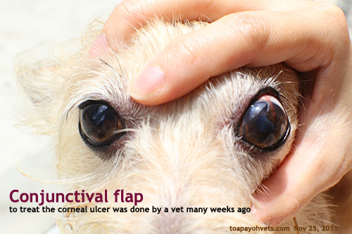
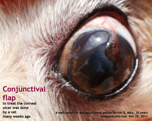
Conjunctival Flap Corneal Ulcer
Refractory Corneal Ulcer Management In Dogs Vet Med At Illinois

Medical And Surgical Management Of Pasteurella Canis Infectious Keratitis Abstract Europe Pmc

The Challenging Approach To A Combined Neurotrophic And Exposure Corneal Ulcer

Corneal Grafts Davies Veterinary Specialists
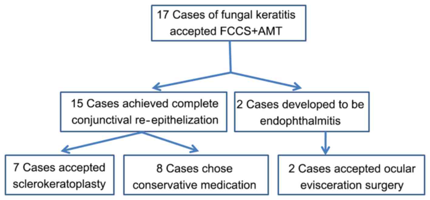
Full Thickness Conjunctival Flap Covering Surgery Combined With Amniotic Membrane Transplantation For Severe Fungal Keratitis
Www Sciencedirect Com Science Article Pii S Pdf Md5 D803de6f519dbad78dd7db298f6aaa16 Pid 1 S2 0 S Main Pdf
A corneal ulcer is an open sore on your cornea, the thin clear layer over your iris (the colored part of your eye) It’s also known as keratitis.

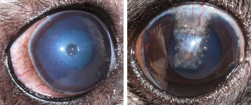
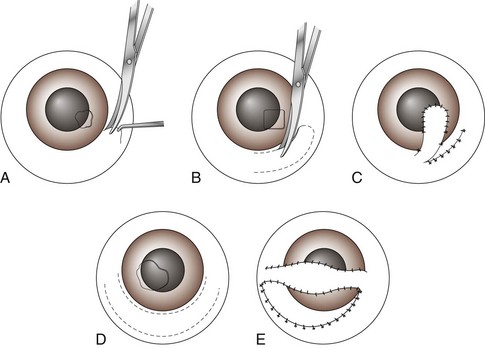
Conjunctival flap corneal ulcer. The purpose of a conjunctival flap is to restore the integrity of a chronically compromised corneal surface, 63,64 and provide metabolic and mechanical support for corneal healing 63,65 They do not add tectonic support and should not be used in very thin or perforated corneas Conjunctival flaps act as biologic patches, conferring a trophic effect because of the nutritional and immunologic supply by its vascular connective tissue. A superficial ulcer with a devitalised epithelial edge where the basal cells are not adhering to the underlying basement membrane will not heal unless the dead tissue is removed, the remaining live tissue encouraged to heal and the area protected. In very deep corneal ulcers or perforations, conjunctival tissue may not be strong enough to provide good structural support to maintain corneal integrity and watertightness Recurrent problems are possible (Figure 7) It also results in significant scarring as the conjunctival tissue is.
The pedicle conjunctival flap can be used either as a thin flap (without Tenon’s capsule) for superficial corneal ulcer, or as a thick flap (with Tenon’s capsule) for deep corneal. Bullous ), a large indolent chronic ulcer, descemetocele, etc, a Gundersen conjunctival flap can often save the eye Futhermore if you have a phthisical eye, first doing a Gundersen flap might allow you later to fit an overlying prothesisTrygve Gundersen MD first. ABSTRACT Thirtythree patients received a conjunctival flap for corneal diseases recalcitrant to medical therapy Twentyfive of them received a total hood flap, and eight a partial conjunctival.
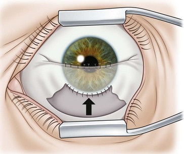
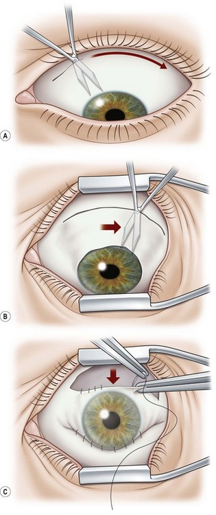
An injection was given below the corneal limbus at the site planned so as to easily cover the ulcer About half a centimetre broad conjunctival flap was created The flap was pulled over the ulcer and fixed by giving three sutures with chronic catgut zero (0) at upper bulbar conjunctiva. Conclusions Conjunctival flap cover surgery is an underused technique Its primary indication is refractory corneal ulcer and corneal perforation, and its second indication is aesthetic with poor visual potential coexisting ocular surface diseases It represents an interesting alternative to more mutilating surgeries. With a truly bad cornea marked diffuse abscess ( ?.
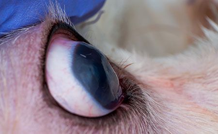
Conjunctival Flap GraftTechnique which sutures a portion of the conjunctiva (pink tissue surrounding the eye) to the cornea for the treatment of deep corneal ulcers Doing so provides an immediate blood supply to the damaged area to speed healing and increase the amount of medication that reaches the area (Figure 15). I) The conjunctival flap covers the affected corneal tissue and prevents tears, proteolytic enzymes, and proinflammatory mediators from reaching the corneal ulcer and causing stromal lysis (11,12) Conjunctival flap surgery in the management of ocular surface disease (Review) MIHAIL ZEMBA1*, ALINACRISTINA STAMATE1*, CALIN PETRU TATARU1,. Conjunctival flap viable option for chronic corneal ulcer Perforated and nonperforated corneal ulcers that have not responded to medical therapies might respond to a selective pedunculated.
With appropriate therapy they typically heal in 3 to 5 days, depending on their initial size Ulcers that persist beyond 5 to 7 days with little improvement despite therapy are considered refractory. The criteria for enrollment in the study were clinical evidence of corneal ulcer that was unresponsive to conventional therapy and the presence of corneal and conjunctival anesthesia. I) The conjunctival flap covers the affected corneal tissue and prevents tears, proteolytic enzymes, and proinflammatory mediators from reaching the corneal ulcer and causing stromal lysis (11,12) Conjunctival flap surgery in the management of ocular surface disease (Review) MIHAIL ZEMBA1*, ALINACRISTINA STAMATE1*, CALIN PETRU TATARU1,.
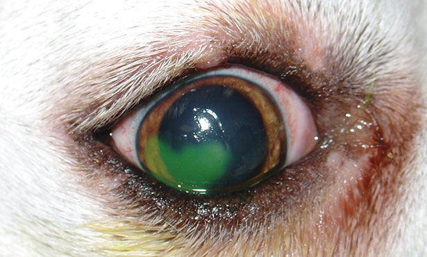
Infectious corneal ulcers are typically caused by secondary bacteria, or less frequently a fungal infection, that triggers corneal stromal destruction Different simultaneous underlying problems such as low corneal sensation, tear film deficiencies and/or eyelid abnormalities can further weaken the cornea and act as contributing factors delaying the corneal healing. Corneal ulcers 1 In our patient, conjunctival flap was performed for resistant fungal corneal ulcer In general, CFs promote wound healing and improve comfort CFs promote healing of refractory. Deep corneal ulcers All corneal wounds are referred to as ‘corneal ulcers’ There are many reasons why a patient may develop a corneal ulcer – including trauma, infection, rubbing from inturned eyelids and dry eye Superficial corneal ulcers are painful but do not pose a threat to the integrity of the globe and are usually treated.
1 Indian J Ophthalmol 19 Jul;30(4)3512 Conjunctival flap in perforated corneal ulcers (A review of cases) Srivastava US, Tyagi RN, Jain AK. Treatment for more than 1 week, but the corneal ulcer Conjunctival flap transplantation is useful for patients withdeepulcers,25Forsuperficialulcers,wewould. Conjunctival flap has been confirmed to be a simple, wellsupported, shortterm treatment for managing corneal perforation or impending perforation in infective corneal ulcers 12–14 However, it is not recommended for the Mooren’s ulcer, a peripheral autoimmunerelated ulcerative corneal diseases.
The corneal ulceration (stromal, descemetocele, or iris prolapse) is covered with the bulbar conjunctival graft that appears most appropriate Postoperative therapy after conjunctival graft placements includes topical, broadspectrum antibiotics, mydriatics, systemic NSAIDs, and systemic antibiotics if the globe was ruptured. Conjunctival flap has been confirmed to be a simple, wellsupported, shortterm treatment for managing corneal perforation or impending perforation in infective corneal ulcers 12–14 However, it is not recommended for the Mooren’s ulcer, a peripheral autoimmunerelated ulcerative corneal diseases. The exposed area of necrotic corneal stroma where the conjunctival flap had dehisced was debrided and the malacic cornea removed using a No 64 Beaver blade and Colibri corneal forceps To ensure removal of all the necrotic stroma, a keratectomy greater than 3/4 of the corneal thickness was carried out by dissection to leave a 30 mm diameter.
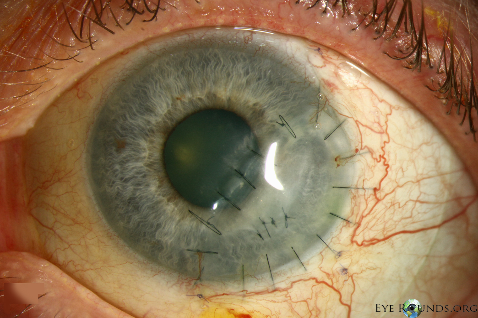
The exposed area of necrotic corneal stroma where the conjunctival flap had dehisced was debrided and the malacic cornea removed using a No 64 Beaver blade and Colibri corneal forceps To ensure removal of all the necrotic stroma, a keratectomy greater than 3/4 of the corneal thickness was carried out by dissection to leave a 30 mm diameter. If the conjunctival flap is being prepared for a perforated corneal ulcer, tissue from the Tenon capsule is mobilized along with the conjunctiva Care is taken to preserve the vascular supply of the flap as much as possible The flap is then secured to the cornea using 100 monofilament nylon sutures. Background Conjunctival flaps are a widely used treatment for numerous corneal ulcers that are caused by microorganismal infections However, whether it can be performed on immunemediated corneal.
A corneal ulcer is a small crater (ulcer) on the front part of the eye, usually resulting from infection Bacteria, viruses, or fungi can cause a corneal ulcer People who wear contact lenses are at higher risk for corneal ulcers because infectious agents may get trapped behind a lens Symptoms of a corneal ulcer include pain, redness,. The top panel is an eye suffering from corneal ulcer and descemetocele (arrow) following a glaucoma shunt procedure and pseudomonas infection After multiple layers of cryopreserved AM, the eye regained vision of /70 seven months later The bottom panel is an eye developing infective keratitis following. Simple ulcers can be treated with prescription eye drops Complex ulcers typically require surgical intervention to remove the damaged or diseased portion of the cornea Our experienced veterinary team provides advanced surgical options for corneal ulcers such as corneal diamond burr debridement and corneal conjunctival flap or graft As veterinary eye specialists, we are able to tailor the proper treatment to your pet.
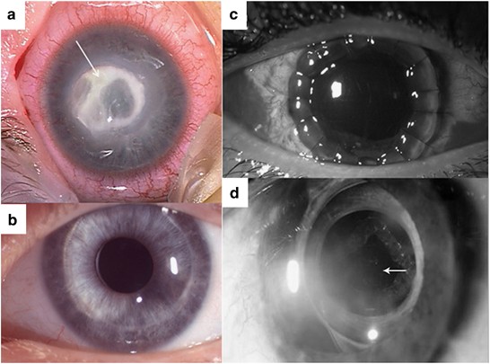
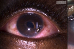
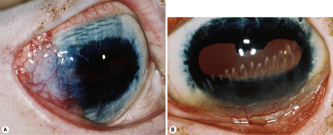
The graft was freely dissected from the ventral palpebral conjunctiva and was sutured to the edge of the deep corneal ulcer with 8/0 Vicryl suture material The eye was covered by a third eyelid flap for two weeks Postoperative management was achieved with topical antibiotics and atropine and systemic nonsteroids. Purpose To describe a case of corneal ulceration associated with Nivolumab use Observations An 80yearold woman treated with Nivolumab for metastatic melanoma developed an intractable corneal ulcer in her left eye, refractory to all therapies including surgery to cover the ulcer with a conjunctival flap until topical prednisolone acetate was tried, which was curative. Conjunctival and episcleral injection are present along with deep vessels in the base of the ulcer A characteristic overhanging central ulcer margin is also seen (B) Total peripheral Mooren’s ulcer with an edematous, opacified central cornea (C) Complete Mooren’s ulcer where a fibrovascular membrane has replaced the corneal stroma.
A corneal ulcer is an ocular emergency that raises highstakes questions about diagnosis and management Four corneal experts provide a guide to diagnostic differentiators and timely treatment, focusing on the types of ulcers most likely to appear in your waiting room When a large corneal ulcer is staring you in the face, time is not on your side. Corneal ulceration, or a break in the corneal epithelium, can occur for a variety of reasons Common etiologies include trauma, entropion, ocular foreign bodies, and dry eye disease Most corneal ulcers are superficial and noninfected;. Yellow dotted circle indicates a corneal ulcer;.
Neurotrophic keratitis ( NK) is a degenerative disease of the cornea caused by damage of the trigeminal nerve, which results in impairment of corneal sensitivity, spontaneous corneal epithelium breakdown, poor corneal healing and development of corneal ulceration, melting and perforation This is because, in addition to the primary sensory role, the nerve also plays a role maintaining the integrity of the cornea by supplying it with trophic factors and regulating tissue metabolism. Conjunctival pedicle grafts The conjunctiva is the pale pink tissue that covers the ‘white’ of the eye It is a thin, strong tissue containing many blood vessels These properties make it a useful graft material for corneal ulcers Conjunctival pedicle grafting is performed with the aid of an operating microscope. The pedicle conjunctival flap can be used either as a thin flap (without Tenon's capsule) for superficial corneal ulcer, or as a thick flap (with Tenon's capsule) for deep corneal ulcer Therefore, the pedicle conjunctival flap can restore ocular surface integrity and provide metabolic and mechanical support for corneal ulcer.
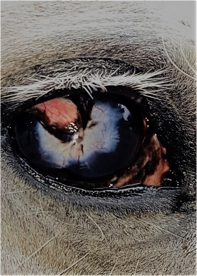
Superficial corneal ulcers treated with medication took days to heal and nine eyes with deep corneal ulcers, treated with conjunctival flap construction, took days to heal All eyes with superficial corneal ulcers recovered within 3 weeks;. The criteria for enrollment in the study were clinical evidence of corneal ulcer that was unresponsive to conventional therapy and the presence of corneal and conjunctival anesthesia. Conjunctival flaps Conjunctival grafts or flaps are used frequently in equine ophthalmology for the clinical management of deep, melting and large corneal ulcers, descemetoceles and perforated corneal ulcers with and without iris prolapse Inappropriate therapy and ulcers.
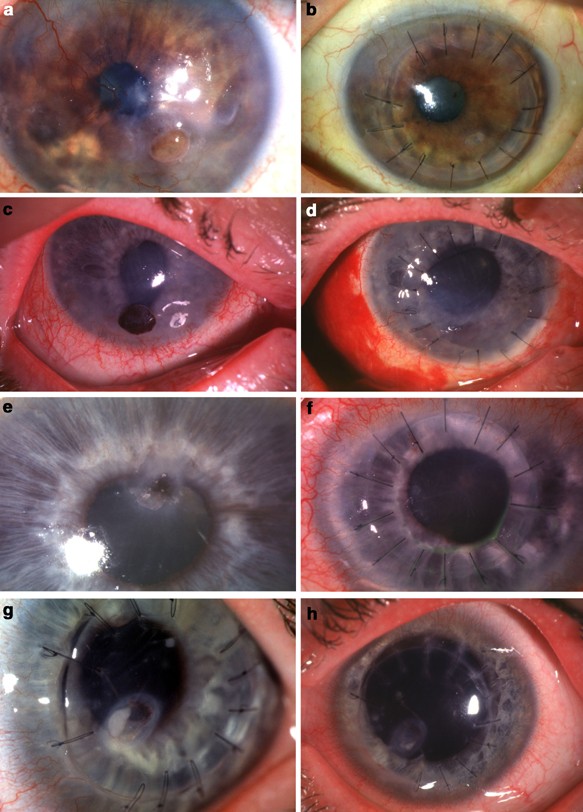
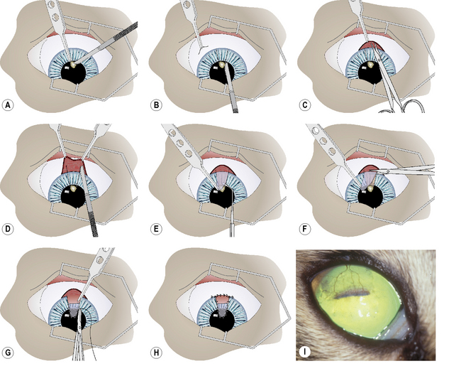
Corneal lesions The conjunctival flap was fully separated toward both ends until it could be pulled to the corneal lesions without tension, and the eyeball could move freely towards all directions Finally, the conjunctival flap was rotated to cover the entire corneal lesions A 10–0 monofilament nylon suture was used to secure the flap with a certain tension (Figure 2F and F’) Any effusion beneath the conjunctival flap should be drive out with a. Conjunctival Flap GraftTechnique which sutures a portion of the conjunctiva (pink tissue surrounding the eye) to the cornea for the treatment of deep corneal ulcers Doing so provides an immediate blood supply to the damaged area to speed healing and increase the amount of medication that reaches the area (Figure 15). Perforation (1×1mm in diameter) with iris prolapse;.
Asterisk indicates a large amount of white purulent secretion emitted from the upper lacrimal punctum b After edgetrimmed PK surgery, a conjunctival flap was used to cover the corneal graft to avoid corneal reinfection. A superficial ulcer with a devitalised epithelial edge where the basal cells are not adhering to the underlying basement membrane will not heal unless the dead tissue is removed, the remaining live tissue encouraged to heal and the area protected. The pedicle conjunctival flap can be used either as a thin flap (without Tenon's capsule) for superficial corneal ulcer, or as a thick flap (with Tenon's capsule) for deep corneal ulcer Therefore, the pedicle conjunctival flap can restore ocular surface integrity and provide metabolic and mechanical support for corneal ulcer.
These were treated with medication alone However, deep corneal ulcers did not. Conjunctival flaps play a less important role in the treatment of perforations than they do in the prevention of progression of corneal melting Nonetheless, in some leaking descemetoceles and. Conjunctival flaps have been used to halt progression of a refractory pseudomonas corneal abscess 10 This is a useful technique in eyes with poor visual potential and a delayed healing rate after both infectious and sterile corneal ulcers A conjunctival flap was used to treat such keratitis occurring in the region of the corneal incisions in a patient after radial keratotomy 11 Flaps have been promoted for treatment of fungal keratitis resistant to medical treatment 12 This approach may.
• Conjunctival pedicle graft The conjunctiva is a thin pink layer which covers the white of the eye It is strong and contains blood vessels, and therefore is a good option to use to repair nearby corneal ulcers A flap of conjunctiva is raised, leaving it attached at it’s base, and it is sutured into the ulcerated area using an. • Conjunctival pedicle graft The conjunctiva is a thin pink layer which covers the white of the eye It is strong and contains blood vessels, and therefore is a good option to use to repair nearby corneal ulcers A flap of conjunctiva is raised, leaving it attached at it’s base, and it is sutured into the ulcerated area using an. When a large corneal ulcer is staring you in the face, time is not on your side “Despite varying etiologies and presentations, as well as dramatically different treatment approaches at times, corneal ulcers have one thing in common the potential to cause devastating loss of vision—often rapidly,” said Sonal S Tuli, MD, associate professor of ophthalmology, director of the cornea and.
Conclusions Conjunctival flap cover surgery is an underused technique Its primary indication is refractory corneal ulcer and corneal perforation, and its second indication is aesthetic with poor visual potential coexisting ocular surface diseases It represents an interesting alternative to more mutilating surgeries. Conjunctival grafts involve taking tissue from the patient' conjunctiva, a pale membrane which covers the white of the eye and includes blood vessels A piece of conjunctiva is moved and rotated so that it covers the ulcer and is then sutured into place Corneal conjunctival transposition (CCT) includes both the conjunctiva and cornea Healthy corneal tissue, with attached conjunctiva, adjacent to the ulcer is moved into the wound, with the conjunctiva providing a functioning blood supply. The Conjunctival Flap or Graft Procedure Conjunctival flap grafting is one option for the treatment of deep corneal ulcers The conjunctiva is the pale pink tissue that covers the “white” of your pet’s eye It is a thin and strong tissue which contains many blood vessels These qualities make it an ideal tissue for grafting purposes Conjunctival flap or grafting surgery is performed under general anesthesia.
Conjunctival flaps have been used to halt progression of a refractory pseudomonas corneal abscess 10 This is a useful technique in eyes with poor visual potential and a delayed healing rate after both infectious and sterile corneal ulcers A conjunctival flap was used to treat such keratitis occurring in the region of the corneal incisions in a patient after radial keratotomy 11 Flaps have been promoted for treatment of fungal keratitis resistant to medical treatment 12 This approach may. When the infection is under control and the ulcerated area needs support conjunctival flap or corneaconjunctival transposition The "Melting" Ulcer The melting refers to the breakdown of the corneal collagen lamellae due to protease, hydrolase and collagenase activities. A corneal ulcer is an open sore that forms on the cornea It’s usually caused by an infection Even small injuries to the eye or erosion caused by wearing contact lenses too long can lead to.
Conjunctival flap (CF) surgery has an acknowledged role in assuring a stable ocular surface and repressing inflammation in compromised corneas or eyes with reduced visual potential In 1958, Gundersen reintroduced and popularized the procedure of using a thin total CF to repair the cornea in a variety of ocular surface diseases Before the refinement of surgical supplies and techniques, almost all perforating corneal injuries were sealed by drawing a hood type flap over the laceration. Results Conjunctival chemosis was observed in 14 out of 16 cases of Pseudomonas aeruginosarelated corneal ulcers, as compared with 6 out of 46 cases caused by other organisms The association between conjunctival chemosis and Pseudomonas aeruginosa is statistically significant, with P value. Cirurgia realizada pelo Dr João Alfredo Kleiner MV, MSc em um cão ShihTzu com ulceração corneana profunda perfurante utilizando a técnica de flap conjuntiv.
Corneal ulcers in dogs are wounds to the outer layer of the eye Learn about the symptoms, diagnosis, and treatment of eye ulcers in dogs Conjunctival flap therapy This treatment involves suturing the dog's third eyelid, located under the lower eyelid, to the inside of the upper eyelid, creating a cover for the cornea. 1 Indian J Ophthalmol 19 Jul;30(4)3512 Conjunctival flap in perforated corneal ulcers (A review of cases) Srivastava US, Tyagi RN, Jain AK. A corneal ulcer is a break in the outer epithelium of the cornea If the outer epithelium alone is breached, the ulcer is superficial If the ulcer is deeper, a certain amount of the underlying stroma is also missing, and the ulcer is deeper Why should I worry about a corneal ulcer?.
Therapeutic Deep Lamellar Keratoplasty For Corneal Perforations Eye

Equine Vision Part Two North Texas Farm And Ranch
Http Aes Amegroups Com Article Download 3699 4445

Key Facts Management Of Deep Corneal Ulcers Semantic Scholar
Canine Dog Veterinary Surgery Anaesthesiaveterinary Surgery Anaesthesia Singapore Toa Payoh Vets Hamster Medicine Surgery Cases Health Sickness Singapore Singapore Toa Payoh Vets

Conjunctival Flaps For Corneal Disease Ento Key
Q Tbn And9gctoywudncljjianeeesklptlrbxqumlkwnxi 4pbj9ur08cdi9h Usqp Cau

Update On Surgical Management Of Corneal Ulceration And Perforation Abstract Europe Pmc

Equine Corneal Ulcer Article Baker Mcveigh
2

Emergency Management Of Corneal Ulcers Veterinary Vision

No Pain No Gain

Understanding Corneal Infection Care

Slit Lamp Photographs Showing The Treatment Course Of Severe Herpes Download Scientific Diagram

A Pain In The Eye Corneal Ulcers In Horses

Corneal Neovascularization Wikipedia

Modified Conjunctival Flap As A Primary Procedure For Nontraumatic Acute Corneal Perforation Sun Yc Kam Jp Shen Tt Tzu Chi Med J
Canine Dog Veterinary Surgery Anaesthesiaveterinary Surgery Anaesthesia Singapore Toa Payoh Vets Hamster Medicine Surgery Cases Health Sickness Singapore Singapore Toa Payoh Vets
Corneal Grafts At Animal Eye Care

Conjunctival Flap Surgery In Iran Conjunctival Flap Cost Iranian Surgery

Corneal Ulcers In Dogs And Catsthe Veterinary Expert Pet Health

Deep Corneal Ulceration And Corneal Grafting Procedures

Management Of Corneal Perforations An Update Deshmukh R Stevenson Lj Vajpayee R Indian J Ophthalmol

Clinical Photographs Illustrating Two Cases Of Mooren S Ulcer That Download Scientific Diagram

Veterinary And Travel Stories 2908 Intern Enucleation
Management Of Inflammatory Corneal Melt Leading To Central Perforation In Children A Retrospective Study And Review Of Literature Eye

Conjunctival Flap Cover Surgery 10 Year Review Yao Annals Of Eye Science

Corneal Ulcer Wikiwand
Q Tbn And9gcsf6urf1qvyyg1bh32hzbldxegtaxugwzffnxqtxcr5dmkxaomr Usqp Cau

Full Text Surgical Therapies For Corneal Perforations 10 Years Of Cases In Opth

Conjunctival Flap Surgery In Iran Conjunctival Flap Cost Iranian Surgery
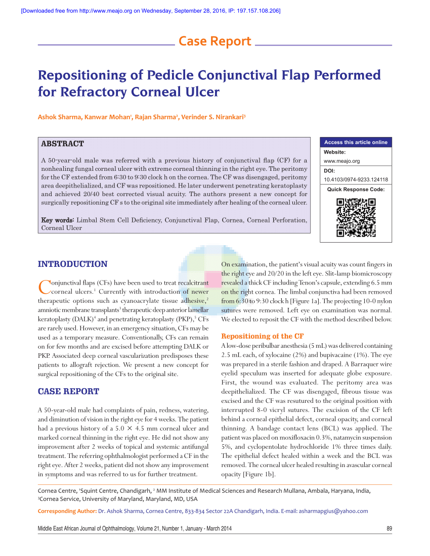
Pdf Repositioning Of Pedicle Conjunctival Flap Performed For Refractory Corneal Ulcer
Peripheral Ulcerative Keratitis Puk
Www Vettimes Co Uk App Uploads Wp Post To Pdf Enhanced Cache 1 Treating Corneal Ulceration In Dogs Part 2 Deep Ulcers Pdf
Why Are Conjunctival Flaps No Longer A Recommended Treatment Quora

Feline Corneal Sequestrum Mspca Angell

The Concept Of Corneal Protection Clinician S Brief

Cureus Treating Mooren S Ulcer Squeezing Water From A Stone

Pin By Cullenwebb Animal Eye Speciali On Eye Diseases Corneal Ulcer Corneal Eyes
Www Agriculturejournals Cz Publicfiles 139 17 Vetmed Pdf

Image Treatment Of Deep Corneal Ulceration With Pedicle Conjunctival Graft Dog Veterinary Manual
Corneal Surgery Clinical Gate

Pdf Rapid Deterioration Of Mooren S Ulcers After Conjunctival Flap A Review Of 2 Cases
Q Tbn And9gcs 8n4e7hdutaj9rdhaonlz2uhimrzk32icgkk0pwybpxijs7s5 Usqp Cau

Ten Tenets One Should Know On Conjunctival Hooding

J Ophthalmol Ukraine 19 4 18 22 Oftalmologicheskij Zhurnal
Why Are Conjunctival Flaps No Longer A Recommended Treatment Quora
Http Www Eyevet Ie Wp Content Uploads 09 01 Deep Corneal Ulcers Pdf

Conjunctival Flap Cover Surgery 10 Year Review Yao Annals Of Eye Science

Case Studies Archives Queenstown Eye Vet

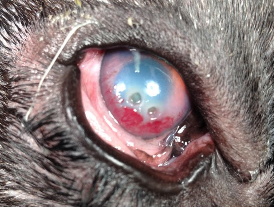
A Cat S Eye With A Conjunctival Pedicle Flap 10 Days After Surgery This Was A Patient Of Dr Cullen S Eyes After Surgery Cats
Corneal Surgery Clinical Gate

Deep Corneal Ulceration And Corneal Grafting Procedures

Community Eye Health Journal Managing Corneal Disease Focus On Suppurative Keratitis
Full Thickness Conjunctival Flap Covering Surgery Combined With Amniotic Membrane Transplantation For Severe Fungal Keratitis

Figure 1 From Rapid Deterioration Of Mooren S Ulcers After Conjunctival Flap A Review Of 2 Cases Semantic Scholar
Rapid Deterioration Of Mooren S Ulcers After Conjunctival Flap A Review Of 2 Cases Bmc Ophthalmology Full Text

Pdf Corneal Ulceration Associated With Nivolumab Use

Djo Digital Journal Of Ophthalmology

Deep Stromal Corneal Ulcers Descemetocele And Iris Prolapse In Animals Emergency Medicine And Critical Care Merck Veterinary Manual

Full Text Neurotrophic Keratitis Current Challenges And Future Prospects Eb

Key Points In Complicated Canine Corneal Ulcers Clinician S Brief

Observations In Ophthalmology Corneal Opacities In Dogs Cats

Corneal Grafts Davies Veterinary Specialists

A Modified Technique Of Keratoleptynsis Letter Box For Treatment Of Canine Corneal Edema Associated With Endothelial Dysfunction Giannikaki Veterinary Ophthalmology Wiley Online Library
Dog Cat Ringworm Treatment Thornleigh Veterinary Hospital

Corneal Grafts Davies Veterinary Specialists

The Challenging Approach To A Combined Neurotrophic And Exposure Corneal Ulcer

Deep Corneal Ulceration And Corneal Grafting Procedures

J Ophthalmol Ukraine 19 4 18 22 Oftalmologicheskij Zhurnal
Tropical Eye Diseases Atlas Last Update 6th June 19 More Coming Tropical Ophthalmology
Conjunctival Flap Covering Combined With Antiviral And Steroid Therapy For Severe Herpes Simplex Virus Necrotizing Stromal Keratitis
Corneal Surgery Clinical Gate
Http S Gridserver Com Wp Content Uploads Eye Surgery In Hot Climates 08 Pdf

Pg Neet Ophthalmology Mcqs Conjunctiva Cornea And Sclera 9
Q Tbn And9gcrtpklkd80qhn H Zf640ywilnz6ty0tuf44upqjs0dpbvhq1au Usqp Cau
Surgery Of The Cornea And Sclera Veterian Key

Extended Care Report After Graft Surgery For A Descemetocoele The Veterinary Nurse
Corneal Surgical Repair

Corneal Ulcers In Dogs Vca Animal Hospital

Mooren S Ulcer Eyewiki

Conjunctival Pedicle Flap Following Removal Of Corneal Sequestrum From A Cat S Eye Corneal Eyes Cats

Canine Pedicle Conjunctival Graft Youtube
Use Of Autologous Fascia Lata Graft To Repair A Complex Corneal Ulcer In A Mare Irish Veterinary Journal Full Text
Surgery Of The Cornea And Sclera Veterian Key
Understanding Canine Ocular Ulcers Dvm 360

Medical And Surgical Management Of Melting Corneal Ulcers Exhibiting Hyperproteinase Activity In The Horse Sciencedirect
Conjunctival Graft Technique In Dogs Vetlexicon Canis From Vetstream Definitive Veterinary Intelligence

Infected Or Stromal Corneal Ulcers Animal Vision Care Surgical Center

Full Text Neurotrophic Keratitis Current Challenges And Future Prospects Eb

Ten Tenets One Should Know On Conjunctival Hooding
Surgery Of The Eye Veterian Key

Sight Saving Op Makes Belle Our December Pet Of The Month 387 Vets
Www Vettimes Co Uk App Uploads Wp Post To Pdf Enhanced Cache 1 Treating Corneal Ulceration In Dogs Part 2 Deep Ulcers Pdf

Equine Corneal Ulcer Article Baker Mcveigh

Corneal Ulcers In Animals Wikipedia
Pet Corneal Ulcers Animal Eye Consultants In Chicago Il

Krishikosh Treatment Of Corneal Ulcers With Prp Solid Buffer Combined With Third Eye Lid Flap And Conjunctival Flap In Dogs A Comparative Study


