Flap Corneal

Full Text Traumatic Corneal Flap Displacement After Laser In Situ Keratomileusis Imcrj
Clinical Efficacy Of Conjunctival Flap Surgery In The Treatment Of Refractory Fungal Keratitis

Late Post Traumatic Flap Dislocation And Macrostriae After Laser In Situ Keratomileusis Sinha R Shekhar H Tinwala S Gangar A Titiyal Js Oman J Ophthalmol

Flap Striae Columbia Ophthalmology
Q Tbn And9gctxxqxjjpvvi4xlgrxpldxjizal5f2ir T5evlptk1oe3wovlvu Usqp Cau

Lasik Flap Versus Surface Prk No Flap Western Laser Eye Associates
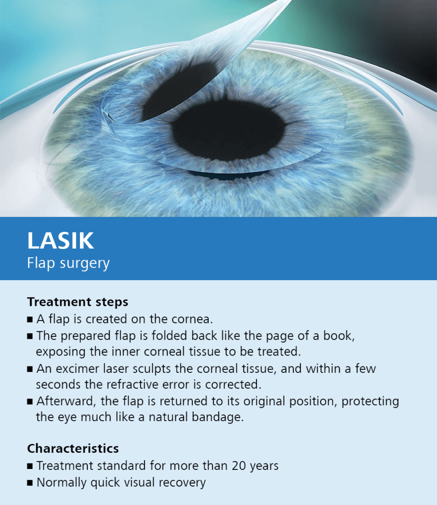
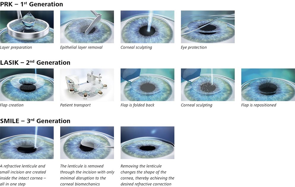
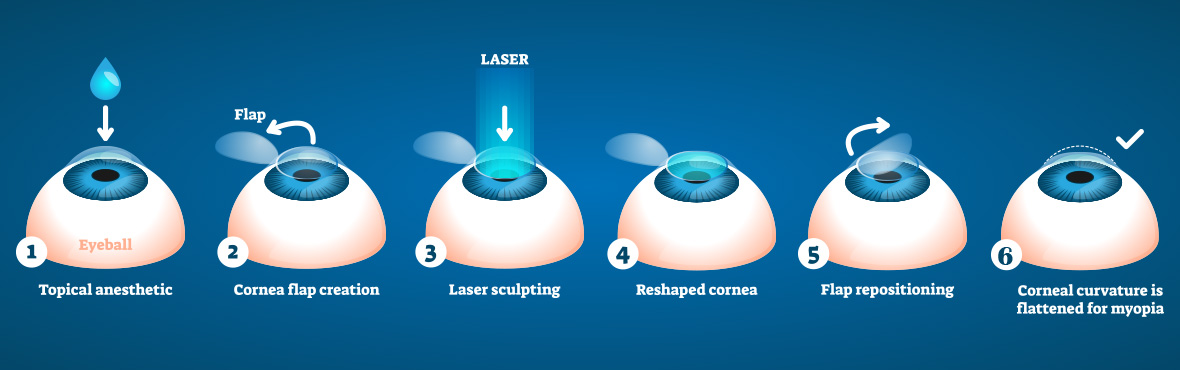
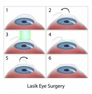
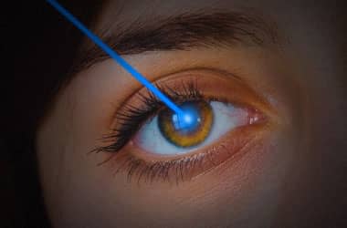
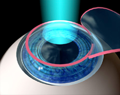
During LASIK eye surgery, the surgeon first administers numbing drops, which take effect in 1015 minutes, and then creates a thin, corneal flap with the laser The surgeon then pulls back the flap to expose the underlying tissue and reshapes the stroma layer of the cornea Then, the laser removes cells according to your prescription.

Flap corneal. Flap vs Lens , , and have to do with the intraocular lens not the corneal flap done for LASIK For the corneal flap dislocation I use 998. The creation of a flap in the outermost layer of the cornea is what distinguishes LASIK from other forms of laser refractive surgery, such as PRK and LASEK This means that flap complications are a potential risk of all forms of LASIK. In October, Alcon’s LenSx femtosecond laser earned a unique distinction by becoming the first laser approved in the United States for LASIK flaps, corneal incisions, anterior capsulotomies and lens fragmentation This approval gives its users more flexibility in how to use the laser, but also presents them with some tough decisions about how to deploy it effectively in their practices, since cataract procedures are usually performed at a site that’s removed from the LASIK suite.
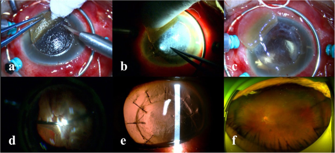
The corneal flap will selfadhere itself naturally in just a few minutes This flap serves as a protective covering for the area of the eye that has been operated on, and thus facilitates rapid healing The corneal flap begins healing immediately after the LASIK procedure In fact, with the use of a LASIK flap, the corneal tissue can be as much as 90% healed within 24 hours. The corneal flap begins healing immediately after the LASIK procedure In fact, with the use of a LASIK flap, the corneal tissue can be as much as 90% healed within 24 hours During the first day or two after surgery, the outer surface of the cornea, known as the epithelium, seals the edges of the corneal flap. Conjunctival flaps have been used to halt progression of a refractory pseudomonas corneal abscess 10 This is a useful technique in eyes with poor visual potential and a delayed healing rate after both infectious and sterile corneal ulcers A conjunctival flap was used to treat such keratitis occurring in the region of the corneal incisions in a patient after radial keratotomy 11 Flaps have been promoted for treatment of fungal keratitis resistant to medical treatment 12 This approach may.
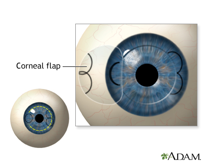
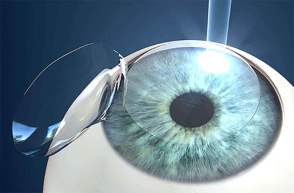
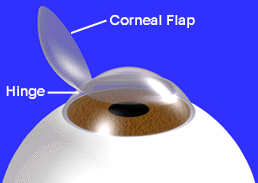
The corneal flap will selfadhere itself naturally in just a few minutes This flap serves as a protective covering for the area of the eye that has been operated on, and thus facilitates rapid healing The corneal flap begins healing immediately after the LASIK procedure In fact, with the use of a LASIK flap, the corneal tissue can be as much as 90% healed within 24 hours. Corneal flap dislocation The corneal flap is altered during every LASIK surgery However, in some cases, this eventually leads to a dislocated corneal flap Although these problems certainly deserve very serious consideration, fortunately there are a number of treatments to remedy them. Creating the Corneal Flap To create the corneal LASIK flap, the LASIK surgeon uses a handheld device or a laser to cut the top layers of corneal tissue at a predetermined depth One edge of the flap is left uncut, forming a hinge Using the hinge, the surgeon folds back the flap to access the underlying corneal tissue.
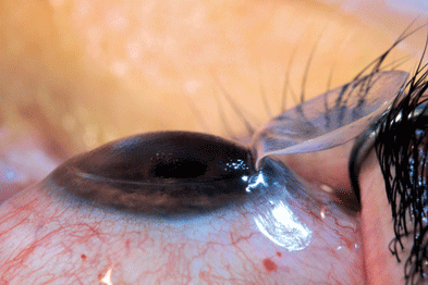
The flap is basically being held into place by osmotic pressure and even excessive eye rubbing could dislodge it After just about any surgery, your body needs time to heal and trauma can disrupt the healing process Fact After a few weeks or so, the outer layer of the cornea has had a chance to regenerate and the flap is more firmly in place. Conjunctival flap grafting is one option for the treatment of deep corneal ulcers The conjunctiva is the pale pink tissue that covers the “white” of your pet’s eye It is a thin and strong tissue which contains many blood vessels These qualities make it an ideal tissue for grafting purposes. A completely detached flap of the cornea Free cap is a rare intraoperative complication of LASIK (laser in situ keratomileusis) Ideally in LASIK, a hinged corneal flap is created that allows eximer laser to be applied on the exposed stromal bed If the hinge of the corneal flap detaches, the flap becomes a free flap/cap.
Pedicle conjunctival flap In small, peripheral perforations, another option is to suture a pedicle conjunctival graft to tamponade the hole Dr Tuli finds this especially useful in nonhealing, neurotrophic ulcers, which are associated with low levels of growth factors in their tear film. In October, Alcon’s LenSx femtosecond laser earned a unique distinction by becoming the first laser approved in the United States for LASIK flaps, corneal incisions, anterior capsulotomies and lens fragmentation This approval gives its users more flexibility in how to use the laser, but also presents them with some tough decisions about how to deploy it effectively in their practices, since cataract procedures are usually performed at a site that’s removed from the LASIK suite. The flap thickness in each sector was calculated by subtracting the mean postflap corneal thickness from the mean preflap corneal thickness In Group 2 (33 eyes), the M2 microkeratome with a 130 microm plate was used to create a superotemporal hinged flap (9 eyes) or a superonasal hinged flap (24 eyes).
Flap vs Lens , , and have to do with the intraocular lens not the corneal flap done for LASIK For the corneal flap dislocation I use 998. Corneal cyanoacrylate gluing is recommended if the cornea appears to be progressively thinning Healing results in approximately 60% of cases Spontaneous perforation should be treated with either. Conjunctival flap grafting is one option for the treatment of deep corneal ulcers The conjunctiva is the pale pink tissue that covers the “white” of your pet’s eye It is a thin and strong tissue which contains many blood vessels These qualities make it an ideal tissue for grafting purposes.
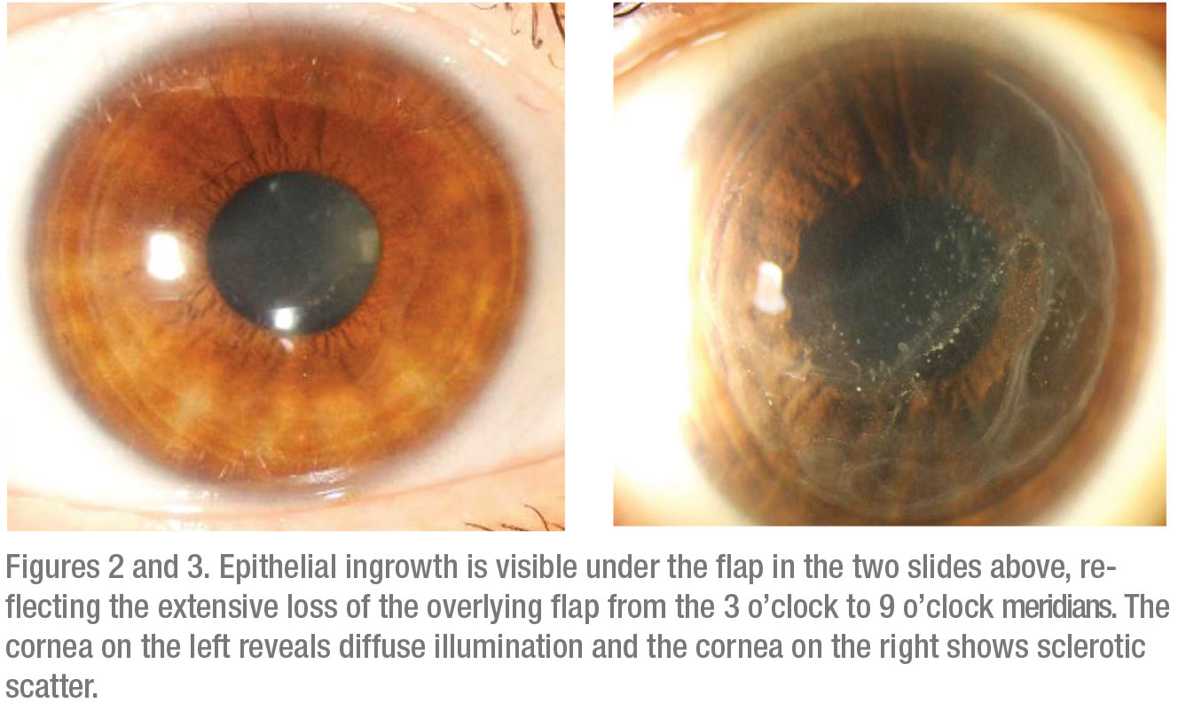
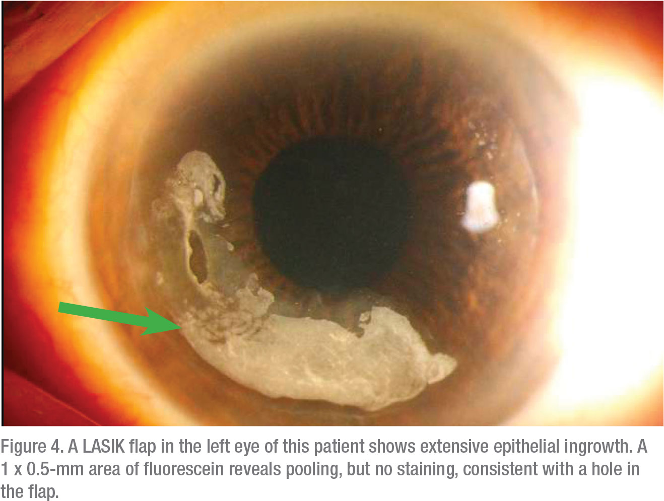
After LASIK, epithelial cells can migrate between the corneal flap and the stromal bed Epithelial ingrowth typically occurs during the first 3 months after surgery, often in the first 1 to 2 weeks The patient may experience a decrease in vision secondary to astigmatic changes from topographic alterations produced by the invading cells. The main indications of therapeutic conjunctival flap cover surgery include unresponsive ulcerative infectious keratitis, persistent corneal epithelial defect, and corneal limbal disease The conventional treatment for acute microbial keratitis is local eye drops. “One of the more common causes for a free flap, especially on an average eye where the cornea is not too flat, is loss of or at least partial loss of suction The eye gets softer, and you are more.
The corneal flap is dislodged and folded Horizontal striae after myopic LASIK are shown here by fluorescein staining Oblique striae are visible by retroillumination. The corneal flaps have free margins about more than 270° without any sutures and anchors except the hinge area, which is attached to the corneal flap smaller than 90°. The corneal flap will selfadhere itself naturally in just a few minutes This flap serves as a protective covering for the area of the eye that has been operated on, and thus facilitates rapid healing The corneal flap begins healing immediately after the LASIK procedure In fact, with the use of a LASIK flap, the corneal tissue can be as much as 90% healed within 24 hours During the first day or two after surgery, the outer surface of the cornea, known as the epithelium, seals the edges.
September 24, 15 The Gundersen conjunctival flap procedure involves the transposition of a thin flap of conjunctiva to cover the cornea for the relief of painful ocular surface disorders or to provide metabolic support for corneal healing This surgical video demonstrates the basic steps of the Gundersen flap procedure in a patient with painful, bullous keratopathy and poor candidacy for keratoplasty. The cornea forms a miniscule scar at the edge of the LASIK flap, which holds the flap in place, but the flap itself does not bond to the underlying cornea Medical research has repeatedly demonstrated that the LASIK flap never heals Peerreviewed medical literature contains numerous reports of flap dislocations many years after LASIK. At Maida Custom Vision, we minimize the risk of flap complications by replacing the traditional microkeratome with an even more accurate, computerguided laser The revolutionary IntraLase® system removes the element of even slight human error, resulting in corneal flaps that have perfectly straight edges, with strong hinges and uniform depths Although using the IntraLase® system to create corneal flaps does not eliminate the possibility of flap complications, it reduces them dramatically.
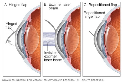
Partialthickness scleral flaps may be dissected with a base at the limbus and then reflected onto the cornea and sutured in place to treat small peripheral corneal perforations To be most. Your cornea steepens, and becomes a cone shape in the bottom portion It usually starts blurring vision during teenage years and worsens during early adulthood. Creating the Corneal Flap To create the corneal LASIK flap, the LASIK surgeon uses a handheld device or a laser to cut the top layers of corneal tissue at a predetermined depth One edge of the flap is left uncut, forming a hinge Using the hinge, the surgeon folds back the flap to access the underlying corneal tissue After the surgeon has reshaped the underlying corneal tissue, the corneal flap is returned to its original position, where it heals naturally.
Flap and cap cuts induced biomechanical weakening in patient corneas The flap caused more weakening than the cap intraoperatively However, biomechanical differences between LASIK and SMILE eyes were similar after removal of tissue and ongoing wound healing J Refract Surg 19;35(5). PRK, or Photorefractive Keratectomy, is usually used for lower degrees of correction It is performed on the surface of the eye LASIK or LaserAssisted InSitu Keratomileusis involves creating a “flap” in the cornea, using the Excimer Laser to reshape the cornea and replacing the flap in its original position. The corneal flap begins healing immediately after the LASIK procedure In fact, with the use of a LASIK flap, the corneal tissue can be as much as 90% healed within 24 hours During the first day or two after surgery, the outer surface of the cornea, known as the epithelium, seals the edges of the corneal flap.
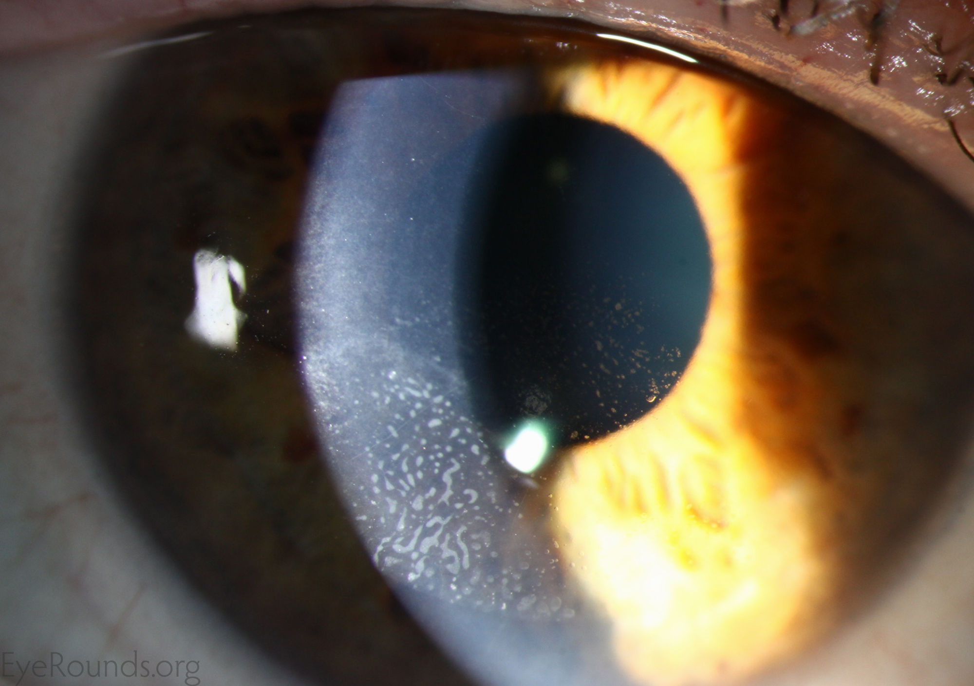
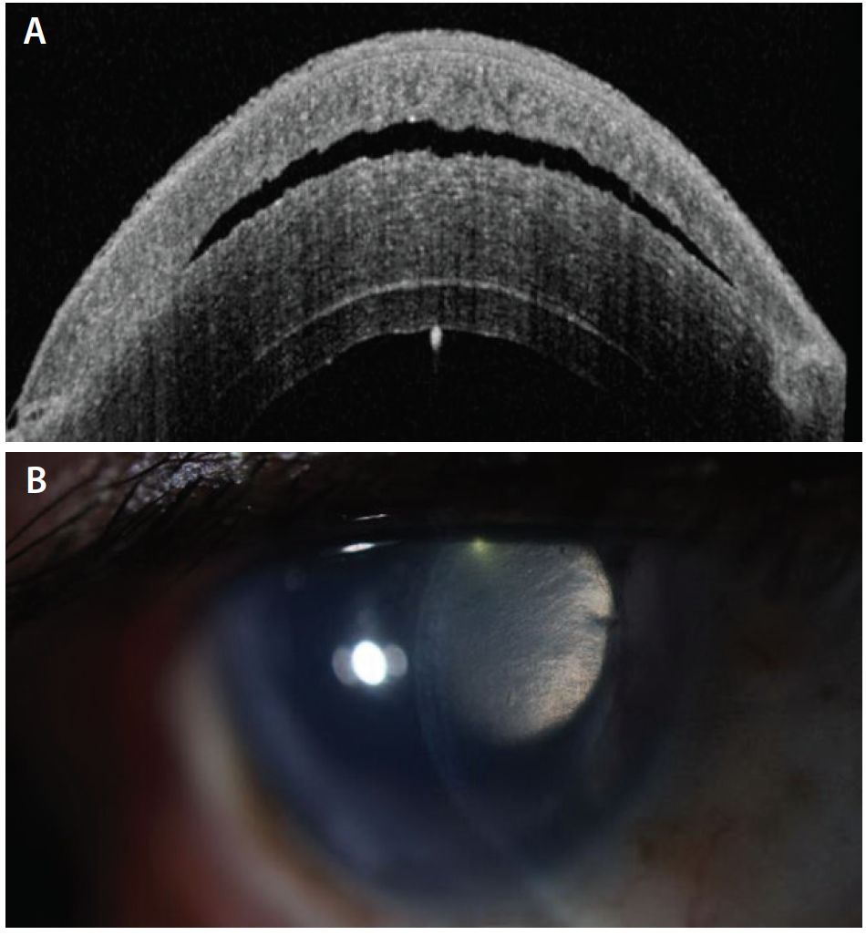
The cornea forms a miniscule scar at the edge of the LASIK flap, which holds the flap in place, but the flap itself does not bond to the underlying cornea Medical research has repeatedly demonstrated that the LASIK flap never heals Peerreviewed medical literature contains numerous reports of flap dislocations many years after LASIK. The healed flap is stable and unlikely to dislodge with eye rubbing Significant eye trauma could potentially dislocate the flap, however, such instances are rare Flap creation making use of the femtosecond laser provides for even greater stability and reduced chances of flap movement or dislocation This has been one of the greatest advances in LASIK surgery. The stroma in the central cornea is 53 microns thinner in the right eye, due to the loss of the flap In the left eye the interface between the LASIK flap performed 16 years earlier and the stromal bed, is not distinctly observable.
Flap striae are defined as small wrinkles or folds in the cornea as a result of LASIK surgery Classification The most common classification of flap striae divides them broadly based on their size Microstriae are microscopic, superficial wrinkles that are generally asymptomatic and often only detectable on microscopic examination of the cornea. During LASIK, a small flap is created in the topmost layer of the corneal, which is known as the epithelium By moving this top layer of the cornea back, a LASIK surgeon can then reshape and recontour the corneal surface and improve the passage of light through the eye Once the corneal reshaping is commplete, the flap is set back down. The Gundersen conjunctival flap procedure involves the transposition of a thin flap of conjunctiva to cover the cornea for the relief of painful ocular surface disorders or to provide metabolic support for corneal healing.
LASIK Complications with Corneas Incomplete or irregular corneal flaps Flaps that are too small or too thin Buttonholes (small holes or tears in the center of the flap) Free caps (flaps without a hinge). The corneal flap heals in different ways at different parts of the flap We’ve broken it down for you here Epithelial layer Your epithelium is the very, very thin layer found on the surface of the cornea It begins healing almost immediately after the flap has been replaced and is the reason that you don’t feel a “line” between the flap and the rest of your eye. The first surgical step in LASIK involves running a surgical blade tangentially across the cornea in order to separate the upper layers from the lower layers and create a “flap” The device the blade is housed in is called a Microkeratome and is analogous to the body of a lathe.
The corneal flap will begin the healing process immediately following the surgery and will be significantly healed in one to three days following the procedure During this time the outer surface of the cornea (also known as the epithelium) will seal the edges of the newly created corneal flap. The flap involves making an incision in the circular strip of the outer corneal tissue, then separating it from the underlying stroma However, one segment of the tissue remains attached by not completing the circle – thus, a small portion of the cornea acts like a hinge Once the tissue is cut to size, the ‘flap’ is complete. The corneal flap will begin the healing process immediately following the surgery and will be significantly healed in one to three days following the procedure During this time the outer surface of the cornea (also known as the epithelium) will seal the edges of the newly created corneal flap.
Find individual business listings for businesses located within the city of Pocatello in Idaho All LASIK Eye Surgery, Surgeons And Centers listings in Pocatello, id Find over 27 million businesses in the United States on The Official Yellow Pages Directory website Find trusted, reliable customer reviews on contractors, restaurants, doctors, movers and more. Corneal flap dislocation The corneal flap is altered during every LASIK surgery However, in some cases, this eventually leads to a dislocated corneal flap Although these problems certainly deserve very serious consideration, fortunately there are a number of treatments to remedy them. If the ingrowth recurs, surgeons get more aggressive with sealing the flap down after the cells are removed “If it recurs, I’ll lift the flap again, remove the epithelial ingrowth, and then use fibrin tissue glue to seal the edge of the flap,” says Dr Manche “My experience with that approach has been quite good.
Process Preoperative procedures Patients wearing soft contact lenses are instructed to stop wearing them 5 to 21 days before Operative procedure A soft corneal suction ring is applied to the eye, holding the eye in place This step in the Postoperative care Patients are usually given a. The benefits of a thinner corneal flap include Faster visual recovery, Higher retinal image quality And shorter treatment times. Creating the Corneal Flap To create the corneal LASIK flap, the LASIK surgeon uses a handheld device or a laser to cut the top layers of corneal tissue at a predetermined depth One edge of the flap is left uncut, forming a hinge Using the hinge, the surgeon folds back the flap to access the underlying corneal tissue After the surgeon has reshaped the underlying corneal tissue, the corneal flap is returned to its original position, where it heals naturally.
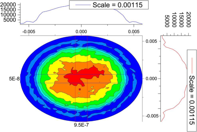
It is known that flap thickness can vary significantly with some mechanical microkeratomes, with standard deviations of 30 µm having been reported depending on the microkeratome used 9 The predictability of this process has a potential for compromising corneal stability in the long term as thicker flaps could more significantly modify the biomechanical integrity of the cornea 10 In this study, we used the femtosecond laser to create flaps at two different depths and measured the impact of. Treatment If identified early, the flap may be relifted, stretched, hydrated and gently smoothed to remove the striae In recalcitrant cases, suture placement at the flap edge has been reported to be successful to remove the striae. A loose flap or a bad flap apposition can dampen the ORA or the surface wave velocity signal Also, 100 and 300 µm in a porcine globe are smaller percentages of total corneal thickness than in human eyes, and the magnitudes of changes seen in the porcine model should not be directly extrapolated to the human case.
“One of the more common causes for a free flap, especially on an average eye where the cornea is not too flat, is loss of or at least partial loss of suction The eye gets softer, and you are more. Automated lamellar keratoplasty (ALK) The surgeon uses an instrument called a microkeratome to cut a thin flap of the corneal tissue The flap is lifted like a hinged door, targeted tissue is removed from the corneal stroma, again with the microkeratome, and then the flap is replaced Laserassisted in situ Keratomileusis (LASIK) The surgeon uses either a microkeratome or a femtosecond laser to cut a flap of the corneal tissue (usually with a thickness of 100–180 micrometres) The flap. Causes of Corneal Perforation 1 Neurotrophic keratitis 2 Peripheral ulcerative keratitis 3 Rosacearelated blepharokeratitis.
Your cornea steepens, and becomes a cone shape in the bottom portion It usually starts blurring vision during teenage years and worsens during early adulthood. If the ingrowth recurs, surgeons get more aggressive with sealing the flap down after the cells are removed “If it recurs, I’ll lift the flap again, remove the epithelial ingrowth, and then use fibrin tissue glue to seal the edge of the flap,” says Dr Manche “My experience with that approach has been quite good. During LASIK surgery, a small flap is created in the frontal, topmost layer of the cornea This layer is called the epithelium Using this epithelial flap, the cornea can be reshaped and recontoured as needed to address refractive errors (ie, myopia/nearsightedness, hyperopia/farsightedness, and astigmatism) What are flap complications?.
To confirm, apply a drop of topical anesthetic to the corneal surface and use a sterile cotton tip applicator to gently wipe the ulcer edge Normal epithelium should not peel away or move Fluorescein stain may also seep underneath the loose epithelium, creating a notable feathered edge to the staining pattern. The stroma in the central cornea is 53 microns thinner in the right eye, due to the loss of the flap In the left eye the interface between the LASIK flap performed 16 years earlier and the stromal bed, is not distinctly observable.

Corneal Graft 3 Months After Undergoing Flap Surgery There Is Perfect Download Scientific Diagram

Pin On Post Refractive Surgery

Lasik Flap Made With Femtosecond Laser Youtube

A Clear Look At The Lasik Flap
Crstoday Cataract Surgery After Lasik Corneal Flap Removal

Recovery Time After Laser Eye Surgery What To Expect Dougherty Laser
Intraoperative Flap Complications In Lasik Prevention And Management Of Free Flaps Springerlink
Q Tbn And9gcsuqogvtezopkkbaabxhlouhhh4fbcqyb9wrltk7rogroagnb8g Usqp Cau
Epithelial Ingrowth Under Lasik Flap
Circular Elliptical Flap Comparison Takes Shape Ophthalmology Times
Before Enhancing Post Lasik Patients
Q Tbn And9gcq 3vda4gl9aihbd2 Slfkmvh8yfq4ul Loc9hsvxwnwo0v Ld2 Usqp Cau
Early Adopter Chronic Sufferer

Eyeworld Bad Memories Of Flap Complications

The Lasik Evolution

Atlas Of Ophthalmology Corneal Perforation And Conjunctiva Flap
Lasik Beverly Hills Lasik Eye Surgery Beverly Hills Gaster Eye

Figure 6 From Corneal Flap Assessment With Rondo Microkeratome In Laser In Situ Keratomileusis Semantic Scholar

A Step By Step Approach To The Diagnosis And Management Of Sands Of Sahara Syndrome Eye News
Modified Deep Anterior Lamellar Dissection For Corneal Opacity During Vitrectomy Case Reports Bmc Ophthalmology Full Text

Hd Corneal Flap Displacement From Eye Trauma After Lasik Em Rap

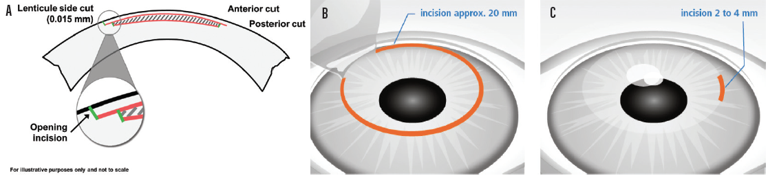
Full Text Verification And Measurement Of The Side Cut Angle Of Corneal Flap In Opth
Lasik Eye Surgery Series Procedure Part 2 Medlineplus Medical Encyclopedia

Before Enhancing Post Lasik Patients
Epithelial Ingrowth Under Lasik Flap

Eyeworld Bad Memories Of Flap Complications

Lasik Complications And Their Management Ento Key

Eyeworld Management Of Rare Post Lasik Complications

How Does The Corneal Flap Heal After Lasek Boozmanhof Rogers

Eyeworld Bad Memories Of Flap Complications
.jpg)
Lasik Flap Dislocation The Flap Never Heals

Lasik Creating The Corneal Flap Service Provider From Bengaluru

Atlas Of Ophthalmology Conjunctival Flap For Corneal Perforation By Professor Chua Chung Nen

Flap Lifting After Corneal Laser Treatment At 2 2 Lj A B And 2 5 Lj Download Scientific Diagram

A Gundersen Conjunctival Flap The Cornea Is Completely Covered By An Download Scientific Diagram

Eyeworld Management Of Rare Post Lasik Complications
Effects Of The Lasik Flap Thickness On Corneal Biomechanical Behavior A Finite Element Analysis Bmc Ophthalmology Full Text

Lasik Flap Surl0001 Stock Eye Images

Repositioning Of Pedicle Conjunctival Flap Performed For Refractory Corneal Ulcer Semantic Scholar
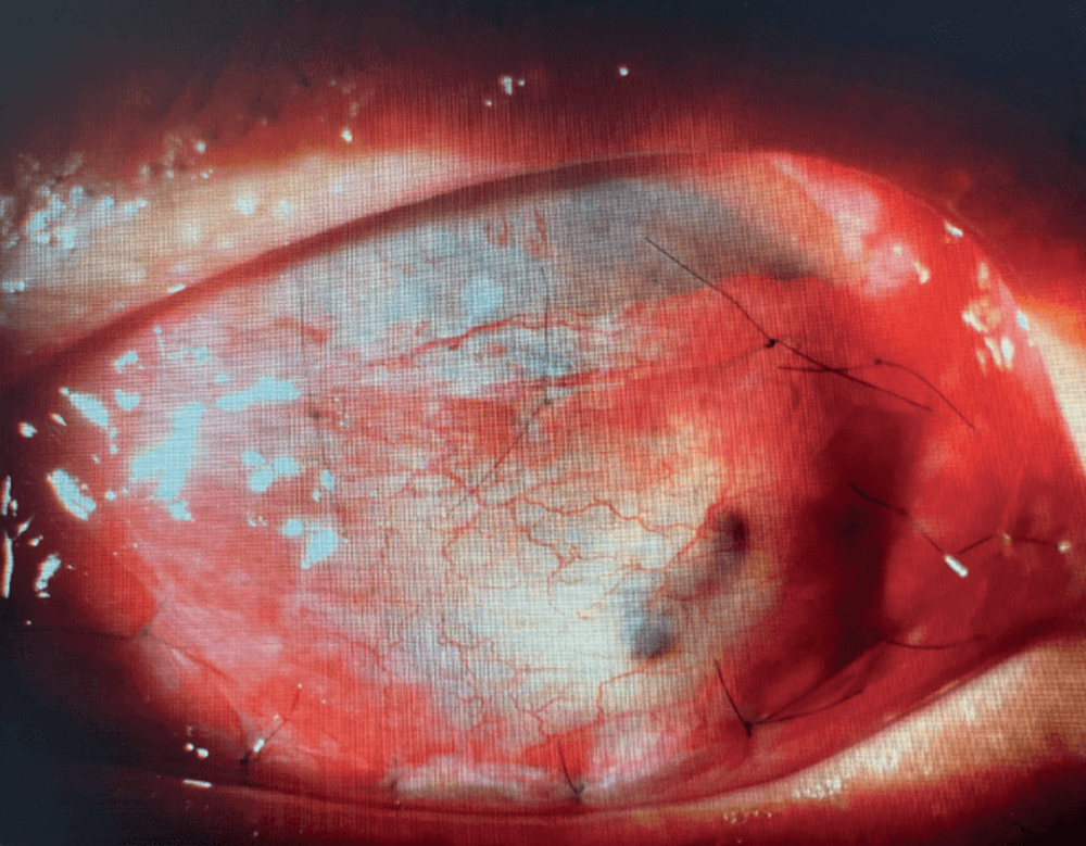
Image Of The Month Gundersen Conjunctival Hooding Flap
Crstoday Visumax From The Patient Perspective
Does Corneal Flap Thickness Matter In Lasik
Before Enhancing Post Lasik Patients
Crsteurope Solo Eye Drop Treatment In Development For Keratoconus

Therapeutic Flap Amputation For Atypical Lasik Flap And Interface Abnormalities

Full Text Verification And Measurement Of The Side Cut Angle Of Corneal Flap In Opth

Biomechanical Corneal Changes Induced By Different Flap Thickness Created By Femtosecond Laser

Posterior Corneal Curvature Changes After Undersurface Ablation Of The Flap And In The Bed Lasik Retreatment Ophthalmology

Long Term Outcomes Of Flap Amputation After Lasik
/ectasia_flap_margin(425).jpg)
Lasik Flap Dislocation The Flap Never Heals
Expert Opinion Laser Eye Surgery Gponline

Schwind Atos The Evolution Of Possibilities Schwind Schwind

The Para Position Of The Corneal Flap In The Left Eye Is Satisfactory 2 Download Scientific Diagram
Why Are Conjunctival Flaps No Longer A Recommended Treatment Quora
Q Tbn And9gcshtyuccybv7vvdxez Ahnisn859qjvndgqzikuakcruwalyzpf Usqp Cau
Epitelizacion De La Interfase Despues De La Tecnica De Lasik Marino Hidalgo Revista Cubana De Oftalmologia

Corneal Flap Creation With Microkeratome Surl00 Eye Illustration Corneal Eye Care
Smile Vs Lasik Laser Eye Surgery

Epithelial Ingrowth Following Laser In Situ Keratomileusis Lasik Prevalence Risk Factors Management And Visual Outcomes Bmj Open Ophthalmology
Procedures Set A New Horizon In Refractive Surgery Ophthalmology Times

Pdf Successful Corneal Flap Replacement Following Complete Traumatic Flap Amputation After Laser Assisted In Situ Keratomileusis Semantic Scholar

Lasik Youtube
Lasik Prk Grosinger Spigelman Grey
Lasik Eye Surgery Mayo Clinic
Lasik Center In Boston Ma Laser Vision Correction Boston Laser

Lasik Enhancement With Epithelial Cells Growing Under Flap Youtube
Full Text Traumatic Corneal Flap Displacement After Laser In Situ Keratomileusis Imcrj
Lasik Vs Prk To Flap Or Not To Flap Better Vision Guide

Successful Treatment Of Severe Wrinkled Corneal Flap After Laser In Situ Keratomileusis With Deionized Water Sciencedirect
Lasik Guider Laser Vision Correction Guide Does The Lasik Flap Ever Heal Completely
Lasik Surgery Explained Definition Procedure Results

Lasik Photo Gallery Sclerallens Com

Repositioning Of Pedicle Conjunctival Flap Performed For Refractory Corneal Ulcer Sharma A Mohan K Sharma R Nirankari Vs Middle East Afr J Ophthalmol
Prk Vs Lasik Eye Care Associates

Patients A Guide To Lasik Prk And Smile Tfos Tear Film Ocular Surface Society
Lasik And Flap Complications Salt Lake City Ut Risks
Femtosecond Laser Assisted Cataract Surgery In A Patient With Traumatic Cataract And Corneal Opacity After Lasik A Case Report Bmc Ophthalmology Full Text

Pin By Cullenwebb Animal Eye Speciali On Eye Diseases Corneal Ulcer Corneal Eyes

Free Cap After Lasik Eyewiki

Figure 2 From Traumatic Flap Dislocation 10 Years After Lasik Case Report And Literature Review Semantic Scholar
Plos One Using Femtosecond Laser To Create Customized Corneal Flaps For Patients With Low And Moderate Refractive Error Differing In Corneal Thickness
/flap_edge(425).jpg)
Lasik Flap Dislocation The Flap Never Heals

Laser Refractive Surgery Lasik
Epitelizacion De La Interfase Despues De La Tecnica De Lasik Marino Hidalgo Revista Cubana De Oftalmologia
Flap Complications From Femtosecond Laser Assisted In Situ Keratomileusis Touchophthalmology

Lifting The Corneal Flap A Alignment Of The Flap B Download Scientific Diagram
Laser Eye Surgery In Zurich Ophthalmologist Dr Selde Zurich

Gundersen Flap In Severe Corneal Edema Youtube
Jcdr Closed Globe Injury Displaced Flap Partial Thickness Laceration Of Cornea
My Eye 6 Days After Lasik Eye Surgery Corneal Flap Healing Red Spots Fading Youtube
.jpg)
Lasik Flap Dislocation The Flap Never Heals

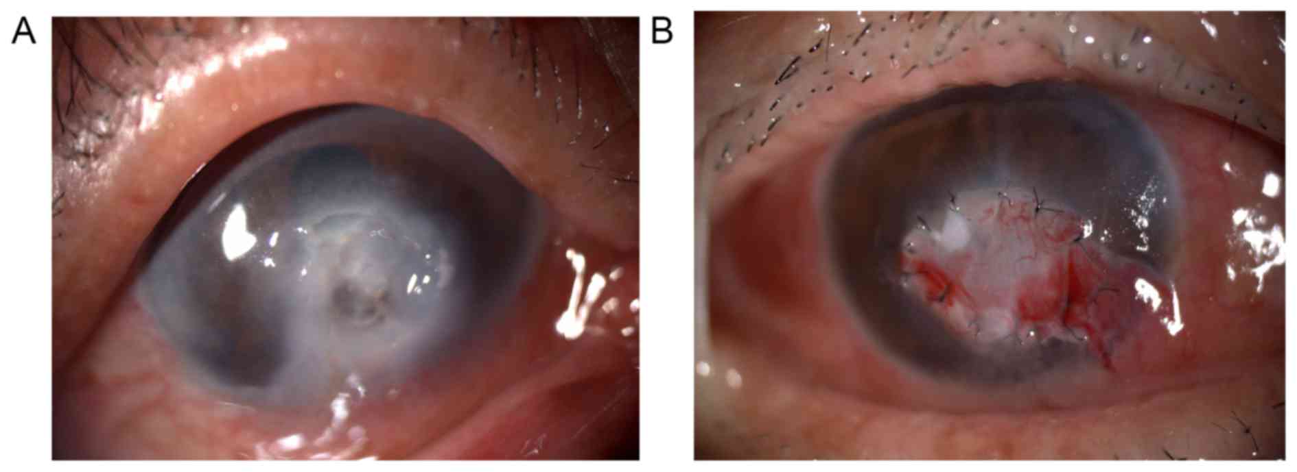
Corneal Flap Dehiscence After Screwdriver Trauma Nejm

Epithelial Ingrowth Columbia Ophthalmology

Welcome To Netra Hospital

Post Lasik Granular Corneal Dystrophy
.jpg)
Lasik Flap Dislocation The Flap Never Heals
/detached-lasik-flap(640).jpg)
Lasik Flap Dislocation The Flap Never Heals
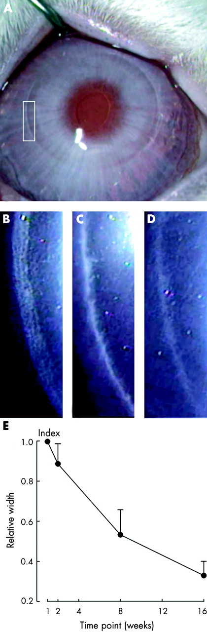
Characterisation Of Corneal Fibrotic Wound Repair At The Lasik Flap Margin British Journal Of Ophthalmology


