Corneal Flap Dog
Third Eyelid Flap Technique In Dogs Vetlexicon Canis From Vetstream Definitive Veterinary Intelligence

Deep Stromal Corneal Ulcers Descemetocele And Iris Prolapse In Animals Emergency Medicine And Critical Care Merck Veterinary Manual
Conjunctival Graft Technique In Dogs Vetlexicon Canis From Vetstream Definitive Veterinary Intelligence

Deep Corneal Ulceration And Corneal Grafting Procedures

Cornea Eye Diseases And Disorders Veterinary Manual

A Modified Technique Of Keratoleptynsis Letter Box For Treatment Of Canine Corneal Edema Associated With Endothelial Dysfunction Giannikaki Veterinary Ophthalmology Wiley Online Library
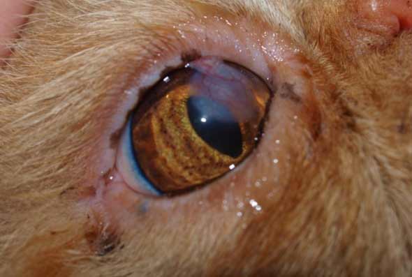
Now the symptoms can vary depending on the type of dystrophy we’re talking about but some of the symptoms might be Discoloration of eyes or rings in the cornea Corneal Lipidosis If your dog has yellowish eyes then it could be corneal lipidosis This can be caused by corneal If your dog has.

Corneal flap dog. The most common type of corneal restructuring graft is the conjunctival pedicle/advancement flap The techniques are suited primarily for restoring the corneal strength (marginal at best though), but is a very poor optical choice These grafts rarely result in clarity (Fig 2). An ulcer is a wound that can occur in different parts of the body However, in this AnimalWised article we are going to focus on explaining the symptoms and treatment of corneal ulcer in dogs, a wound that occurs in the cornea of a dog Due to its location it will always require veterinary intervention, since leaving it untreated can result in significant damage to the eye that even leads to. Understanding Corneal Endothelial Degeneration Corneal Endothelial Degeneration (CED) is a degenerative condition in dogs that affects the clarity of the cornea This agerelated disease can result in blindness and severe ocular pain from secondary complications The cornea is the clear window at the front of the eye.
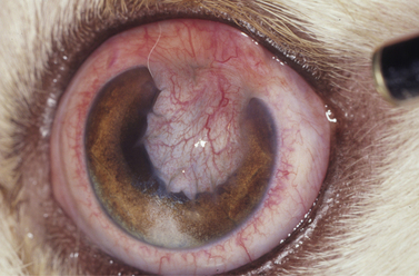
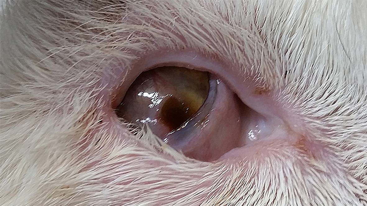
My dog's conjunctival flap surgery?. Daniel M Dorbandt, Phillip A Moore, Kathern E Myrna, Outcome of conjunctival flap repair for corneal defects with and without an acellular submucosa implant in 73 canine eyes, Veterinary Ophthalmology, /vop, 18, 2, (), (14). Diffuse corneal edema in a dog with a penetrating corneal cat claw injury and secondary uveitis The wound from the cat claw can be seen on the ventromedial paraxial cornea Treatment involves emergency surgical support—either conjunctival grafting or placement of a third eyelid flap 4,5 FIGURE 3 Feline acute bullous keratopathy Note.
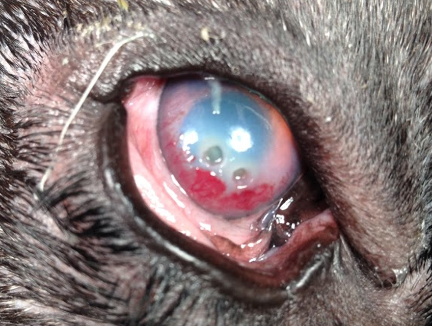
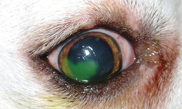
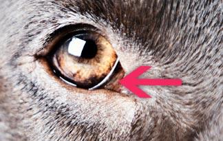
When deep ulcers occur, the membrane that covers the inner surface of the cornea may protrude through the cornea, or the ulcer can create a fullthickness hole in the cornea In dogs, most ulcers are caused by injury, such as nail scratches, foreign objects in the eye, or chemicals that enter the eye. Canine Corneal Abrasions Symptoms of Corneal Abrasions Chip may paw his eye or blink a lot if he is bothered by a corneal abrasion Other Causes of Corneal Abrasions It's not difficult for Chip to get a corneal abrasion A poke or scrape, a foreign body, or Treating a Corneal Abrasion The. Common signs of corneal ulcers in dogs and cats include squinting, redness in the sclera (the part of the eye that is normally white), abnormal discharge from the eye – either watery or mucousy – and sometimes cloudiness or haziness to the eye itself.
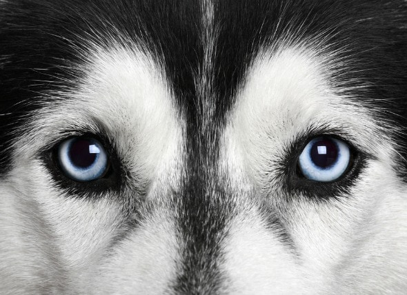
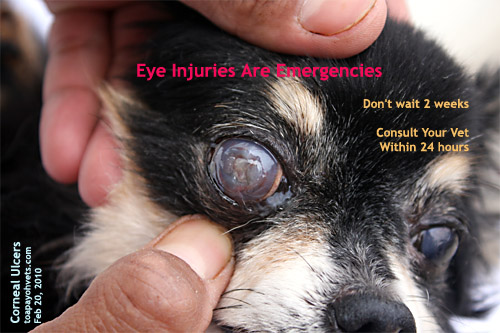
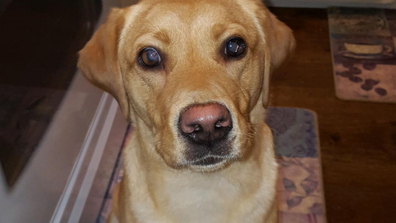
The hallmark sign of corneal endothelial dystrophy in dogs is eyes with a blue or foggy appearance As the disease progresses, patients experience ocular discomfort and pain Your pet may also avoid bright light or show signs of visual discomfort when outdoors. The cornea is the transparent, shiny membrane that makes up the front of the eyeball With a corneal ulcer, fluid is absorbed from the tears into the stroma, giving a cloudy appearance to the eye The most common cause of a corneal ulcer is trauma Less common causes of corneal ulcers include bacterial infections, viral infections, and other diseases. Superficial keratectomy and 360° conjunctival flap for bullous keratopathy in a dog A case reportpdf (Milvago chimachima) treated with a modified third eyelid flap 1 tion of the cornea.
Purpose To evaluate the efficacy of superficial keratectomy and conjunctival advancement hood flap (SKCAHF) for the treatment of bullous keratopathy in canine patients Methods Nine dogs (12 eyes) diagnosed with progressive corneal edema underwent superficial keratectomy followed by placement of conjunctival advancement hood flaps. A corneal ulcer is the deep erosion of the eye’s third layer, which results in a cloudy appearance and pain for your pet It is commonly caused by trauma – either via the eyelashes rubbing against the eye (entropion), via a cat scratch or via contact with a sharp object. Symptoms of Corneal Dystrophy in Dogs White or grayish cloud in the center of the eye Irritated eyes.
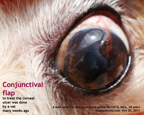
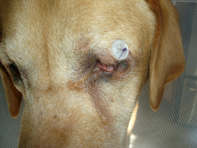
Option to use to repair nearby corneal ulcers A flap of conjunctiva is raised, leaving it attached at it’s base, and it is sutured into the ulcerated area using an In the immediate postoperative period, the dog needs to be watched carefully Your pet may be a little groggy due to the anaesthetic that was required It is not unusual for the. In all dogs, corneal dystrophy is caused by a genetic disturbance in how fat is metabolized The result is a white or gray clouding of the eye It generally starts in one eye but always affects both In most breeds, it does not cause discomfort or blindness In a smaller list of breeds the disorder is more progressive and can lead to more. Endothelial corneal dystrophy may be treated by using contact lenses over your dog's eyes Epithelial corneal tags may be removed, if present Another possible treatment for endothelial corneal dystrophy is flap surgery of the conjunctiva (the lining of the eyeball and the back surface of the lids).
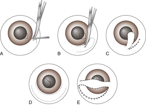
To report and compare the success rate of a conjunctival pedicle flap (CPF ) alone vs a CPF with an underlying acellular submucosa implant for the repair of deep or perforating corneal wounds in dogs Procedures Records of 69 dogs (73 eyes) receiving a CPF with or without an acellular submucosa implant between 04 and 12 were reviewed. Young dogs can also develop ectopic cilia, or abnormal hairs that protrude from the conjunctiva lining the eyelid When the animal blinks, these cilia contact the cornea and create ulceration Surgery, typically under an operating microscope, is required to remove the ectopic cilia and allow the ulcer to heal. The area is either outlined with a corneal trephine of appropriate size or with a 64 Beaver blade, and then it is undermined with a corneal dissector The flap of cornea, usually 150 to 0 µm thick, is then removed, and any attachments are trimmed as necessary Following the keratectomy, either a contact lens or third eyelid flap may be placed.
Treating corneal ulceration in dogs part 2 deep ulcers Author Mateusz Jaksz, Claudia Busse Corneal sampling for cytology and, ideally, culture and sensitivity is recommended for the This type of flap is easy to perform, but covers the whole cornea and, therefore, completely impedes vision and precludes adequate monitoring of the. Treating corneal ulceration in dogs part 2 deep ulcers Author Mateusz Jaksz, Claudia Busse Categories Companion animal, Vets Date March , 17 Deep corneal ulcers are defined by a defect of the epithelium and stromal loss that may extend as far as the Descemet’s membrane Figure 1a Deep corneal ulcer in two pugs, frontal view. Option to use to repair nearby corneal ulcers A flap of conjunctiva is raised, leaving it attached at it’s base, and it is sutured into the ulcerated area using an In the immediate postoperative period, the dog needs to be watched carefully Your pet may be a little groggy due to the anaesthetic that was required It is not unusual for the.
The latter is “almost like a rubbery piece of tissue that, in some ways, is even a little easier to dissect than a partialthickness corneal piece,” said Dr Karp Irradiated sterile cornea also has a long shelf life and is applicable to nonendothelial procedures 2 It’s also possible to temporarily transplant other types of banked tissue. In dogs and cats damage to the cornea is a very common occurrence, because they lead with their head and often hit things that can bump or damage the eye Dogs can also get corneal ulcers from roughhousing or rubbing their head on the ground Cats sometimes get corneal ulcers from fighting with other cats. Corneal ulcers are eye problems resulting from a loss in the tissue layers of your pet’s cornea, or lens This eye problem results in a pain and can impact their vision There are many potential causes for corneal ulcers such as trauma, disease, foreign body, or infection.
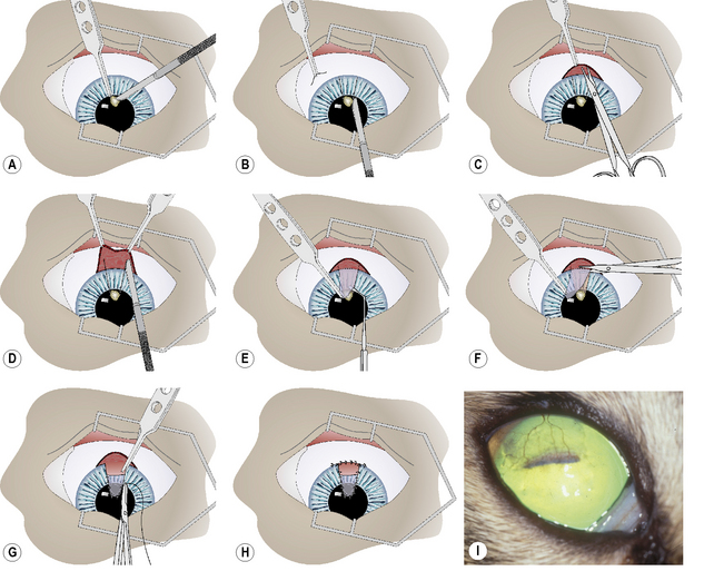
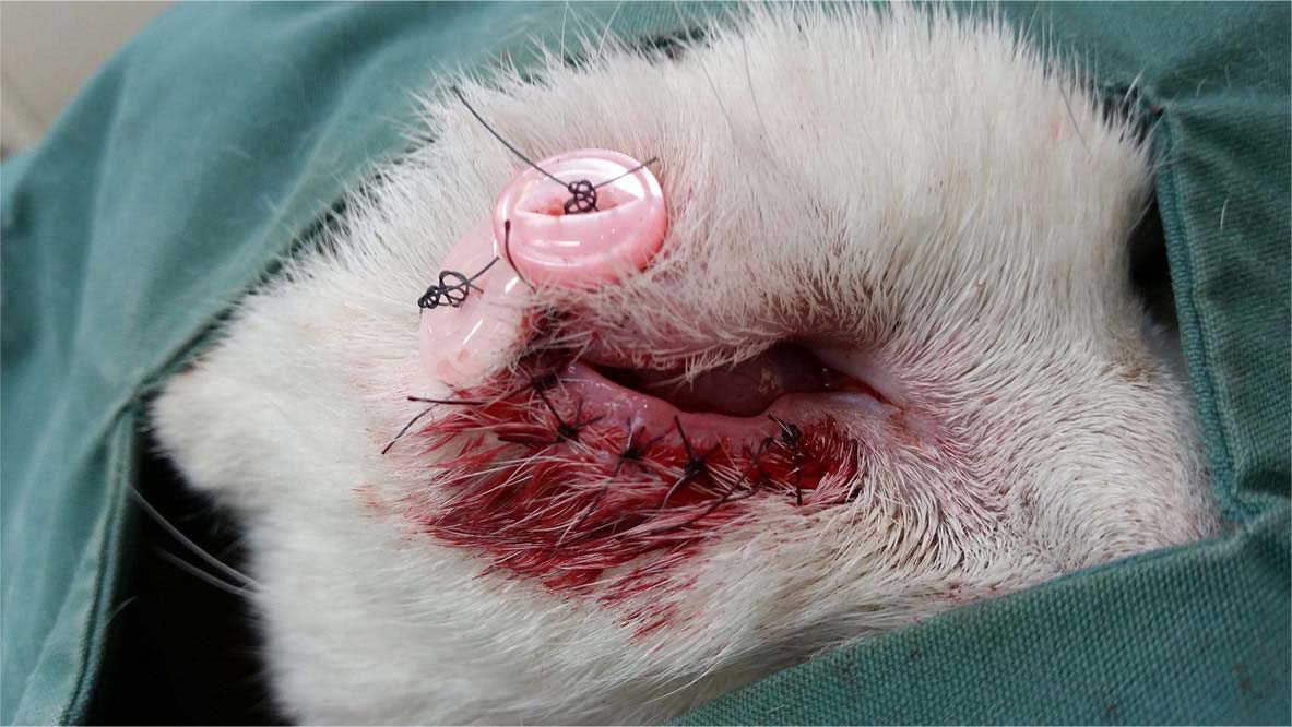
Conjunctival flap cut scenes 犬のデスメ膜瘤保護のための結膜フラップ作成シーン part 1 は結膜弁の切り出しシーン。. An ulcer is a wound that can occur in different parts of the body However, in this AnimalWised article we are going to focus on explaining the symptoms and treatment of corneal ulcer in dogs, a wound that occurs in the cornea of a dog Due to its location it will always require veterinary intervention, since leaving it untreated can result in significant damage to the eye that even leads to. In this VETgirl online veterinary continuing education video, we discuss the use and placement of a 3rd eyelid flap in your veterinary patientA third eyelid flap is an easy and quick procedure, and is a very good option for certain cases The 3rd eyelid can serve as tectonic support and protection for severe, melting corneal ulcers or large corneal perforations when a conjunctival graft is.
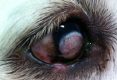
Topical antibiotic use was terminated 13 days sooner ( P ≤ 001) in dogs with an acellular submucosa implant The combined success rate of all corneal wounds was 93% with success rate of corneal perforations, descemetoceles, and deep stromal wounds being %, 95%, and 100%, respectively. Thermal keratoplasty (a technique for reshaping the cornea) and Gunderson flap (replacing the damaged section of cornea with a section or “flap” of the patient’s own conjunctiva) procedures for corneal edema (swelling) associated with corneal endothelial degeneration (a condition in dogs that affects corneal clarity). Nonhealing Superficial Corneal Ulcers in Dogs by Daniel Biros, DVM, DACVO Sometimes loose flaps of epithelial sheets or fragments of epithelia from the wound edges are seen freely hanging from the ulcer’s edge, misguided and unsuccessful attempts at wound healing Most cases are unilateral, but bilateral ulcers may present.
Nonhealing Superficial Corneal Ulcers in Dogs by Daniel Biros, DVM, DACVO Sometimes loose flaps of epithelial sheets or fragments of epithelia from the wound edges are seen freely hanging from the ulcer’s edge, misguided and unsuccessful attempts at wound healing Most cases are unilateral, but bilateral ulcers may present. The latter is “almost like a rubbery piece of tissue that, in some ways, is even a little easier to dissect than a partialthickness corneal piece,” said Dr Karp Irradiated sterile cornea also has a long shelf life and is applicable to nonendothelial procedures 2 It’s also possible to temporarily transplant other types of banked tissue. Corneal abrasions are one of the most common forms of eye injury In some cases, they are caused by the direct impact of a sharp object, such as a pencil, staple, nail or sewing pin They also can be caused by small, airborne particles, such as dust, sand or flying debris from soldering, woodworking or weed trimming.
The dog in this report undergoing corneal débridement with a diamond burr to treat a spontaneous chronic corneal epithelial defect in the left eye The dog's therapy of topical 1% morphine solution administered three times a day for pain management was reinstituted and the antibiotic ophthalmic ointment was continued. Corneal ulcer is a condition that may affect dogs of all ages Chronic ulcers may be more common in middle aged and senior dogs The ulcers may be caused by injuries, bacterial, viral or fungal infections Treatment should be applied, as these ulcers may affect the vision of the pet and may also cause blindness. Nonhealing Superficial Corneal Ulcers in Dogs by Daniel Biros, DVM, DACVO Sometimes loose flaps of epithelial sheets or fragments of epithelia from the wound edges are seen freely hanging from the ulcer’s edge, misguided and unsuccessful attempts at wound healing Most cases are unilateral, but bilateral ulcers may present.
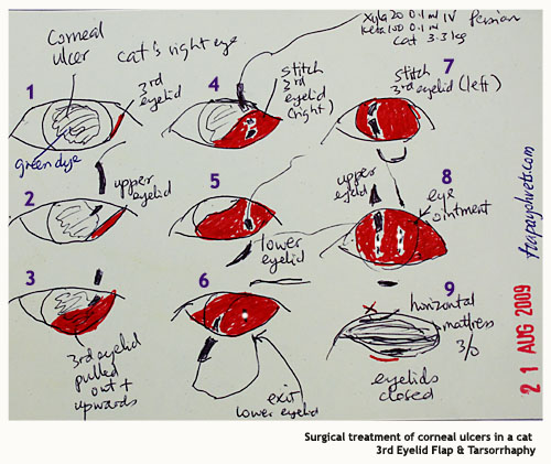
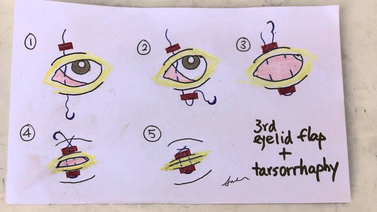
I told the senior vet "I do treat corneal ulcers using the 3rd eyelid flap method and do stitch up corneas if necessary" The senior vet told me "Tarsorrhapy is very effective in corneal ulcer cases Even in cases where the eyeball collapses No need to stitch up the cornea* Just sew up the two eyelids and the outcome is very good. Syndromes of very slowhealing and recurrent shallow ulcers occur in dogs, especially in older animals They may be due to a membrane disease causing faulty attachment of the thin layer of cells lining the cornea or due to a virus called herpesvirus. Gundersen Conjunctival Flap Contributors Jesse M Vislisel, MD and Mark A Greiner, MD September 24, 15 The Gundersen conjunctival flap procedure involves the transposition of a thin flap of conjunctiva to cover the cornea for the relief of painful ocular surface disorders or to provide metabolic support for corneal healing.
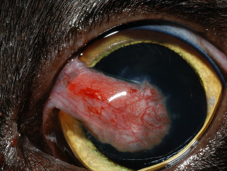
She had it done because of a corneal ulcer and the vet wanted a blood supply to get in there i'm just not sure what to expect after the surgery i've called the vet with what i thought were changes but they said i only need to worry if she's squinting or keeping her eye. Corneal ulcers are a common problem in pets, and we offer a variety of treatments, including conjunctive flap grafting surgery The Conjunctival Flap or Graft Procedure Conjunctival flap grafting is one option for the treatment of deep corneal ulcers The conjunctiva is the pale pink tissue that covers the “white” of your pet’s eye. In this VETgirl online veterinary continuing education video, we discuss the use and placement of a 3rd eyelid flap in your veterinary patientA third eyelid flap is an easy and quick procedure, and is a very good option for certain cases The 3rd eyelid can serve as tectonic support and protection for severe, melting corneal ulcers or large corneal perforations when a conjunctival graft is.
Some possibilities include Conjunctival flap therapy This treatment involves suturing the dog's third eyelid, located under the lower eyelid, to Keratotomy This is a surgery during which the cornea involved in the ulcer is pierced or cut to encourage a new layer Soft contact lenses. Conjunctival flaps may be used as adjuncts to lamellar or fullthickness grafts in melting disorders to stabilize the surface and prevent lytic destruction of the grafts 16 Despite their reinforcing ability, conjunctival flaps should not be considered sole management for corneal perforations A flap will help the anterior chamber to reform. In this VETgirl online veterinary continuing education video, we discuss the use and placement of a 3rd eyelid flap in your veterinary patientA third eyelid flap is an easy and quick procedure, and is a very good option for certain cases The 3rd eyelid can serve as tectonic support and protection for severe, melting corneal ulcers or large corneal perforations when a conjunctival graft is.
Third eyelid flap sutured across to protect the cornea/allow the ulcer to heal (with surgery to the lower eyelid to correct the entropion that was the cause of the ulcer) Treatment options Some cases can be treated with eye drops/ointment containing antibiotics that will both prevent infection and act as a lubricant, to reduce some of the. THE TECHNIQUE of using a conjunctival flap for the treatment of chronic corneal ulceration was described by Gunderson 1 in the late 1950s and became a standard surgical procedure There are some drawbacks to this procedure, however 1,2 During the past years, progress in microsurgical technique, aided by the microscope and fine surgical tools, has enabled us to use a selective pedunculated. Examples of other diseases include epithelial dystrophy a weakening of the cornea which can be inherited in breeds such as Boxer Dogs drying of the cornea due to decreased tear production, called keratoconjunctivitis sicca (KCS or "dye eye") endocrine diseases such as diabetes mellitus, Cushing's.
Large Replacement Dog Door Flap Compatible with PetSafe Freedom Doggie Doors PAC Measures 10 1/8" x 16 7/8" Made from flexible, durable, weather resistant materials Doggie Door Flap 45 out of 5 stars 1,928 $2699 $ 26 99 ($2699/Count) Get it as soon as Fri, Jan 22. TyBo underwent a conjunctival flap procedure (or ‘conjunctival flap grafting’), which is a more common option for severe or deep corneal ulcers Essentially, the conjunctiva is the pale pink tissue that covers the “white” of your pet’s eye, and through surgery, a specialist relocates and grafts a portion of this tissue to cover the. If the dog is cooperative a grid keratotomy can be done with a nurse holding the dog I use a 25 gauge needle and make linear incisions into the superficial corneal stroma Do this in lines say dorsoventral over the debrided area, then make grids by making linear incisions at 90 degrees to the first set of lines.
Some breeds of dogs, such as Boxers, have genetic corneal abnormalities predisposing them to ulcers Corneal ulcers are painful Shutting the eye or excessive blinking or spasm of the eyelids and a watery discharge are common signs The pet may avoid bright light. The aim of this study was to report a case of BK in a dog and the complete recovery of the ocular structure and visual function, with a third eyelid flap associated with the use of autologous blood serum topicallyCase A 2yearold Shih Tzu male dog, weighing 43 kg, with recurrent bilateral eye discomfort was brought to Ophthalmologist Veterinarian Assistance. Thermal keratoplasty (a technique for reshaping the cornea) and Gunderson flap (replacing the damaged section of cornea with a section or “flap” of the patient’s own conjunctiva) procedures for corneal edema (swelling) associated with corneal endothelial degeneration (a condition in dogs that affects corneal clarity).

Lasik Flap Lift And Irrigation After Dog Scratch Injury Youtube

3 Ways To Treat Canine Corneal Ulcers Wikihow
Www Vettimes Co Uk App Uploads Wp Post To Pdf Enhanced Cache 1 Treating Corneal Ulceration In Dogs Part 2 Deep Ulcers Pdf
Canine Dog Veterinary Surgery Anaesthesiaveterinary Surgery Anaesthesia Singapore Toa Payoh Vets Hamster Medicine Surgery Cases Health Sickness Singapore Singapore Toa Payoh Vets

Full Text Traumatic Corneal Flap Displacement After Laser In Situ Keratomileusis Imcrj

Treating A Corneal Ulcer In A Bug Eyed Dog Youtube

The Concept Of Corneal Protection Clinician S Brief
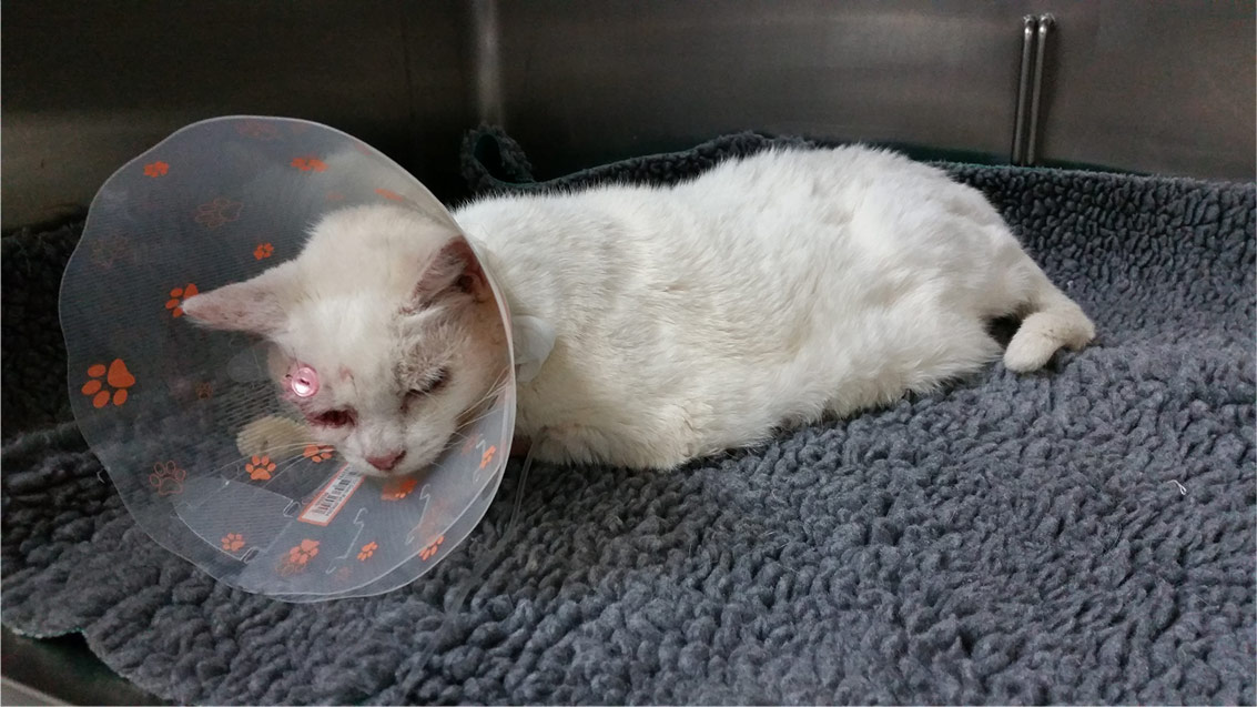
My Dog Had A Third Eye Lid Flap Today Around 3 00pm I Picked Her Up St 5 00 She Has The Cone Collar That Is Really
Q Tbn And9gcq3tc2qknuxaz0e53r4piiuf6p8ttpw3li2e4joqgjaqrn3ielj Usqp Cau

How To Place A 3rd Eyelid Flap Into A Dog Or Cat Vetgirl Vet Ce Video

Clinical Approach To The Canine Red Eye Today S Veterinary Practice

Veterinary And Travel Stories 3229 Intern Tarsorrhaphy And 3rd Eyelid Flap Procedures For Eye Corneal Ulcerations In Toa Payoh Vets

Corneal Ulcers

Eye Problems In Dogs And Cats Treatment Of Corneal Ulcers The Hi Lo

Deep Corneal Ulceration And Corneal Grafting Procedures
Understanding Canine Ocular Ulcers Dvm 360
Pet Corneal Ulcers Animal Eye Consultants In Chicago Il

Pdf Incidence Of Corneal Ulcer In Dogs A Retrospective Study Semantic Scholar

The Concept Of Corneal Protection Clinician S Brief
Refractory Corneal Ulcer Management In Dogs Vet Med At Illinois
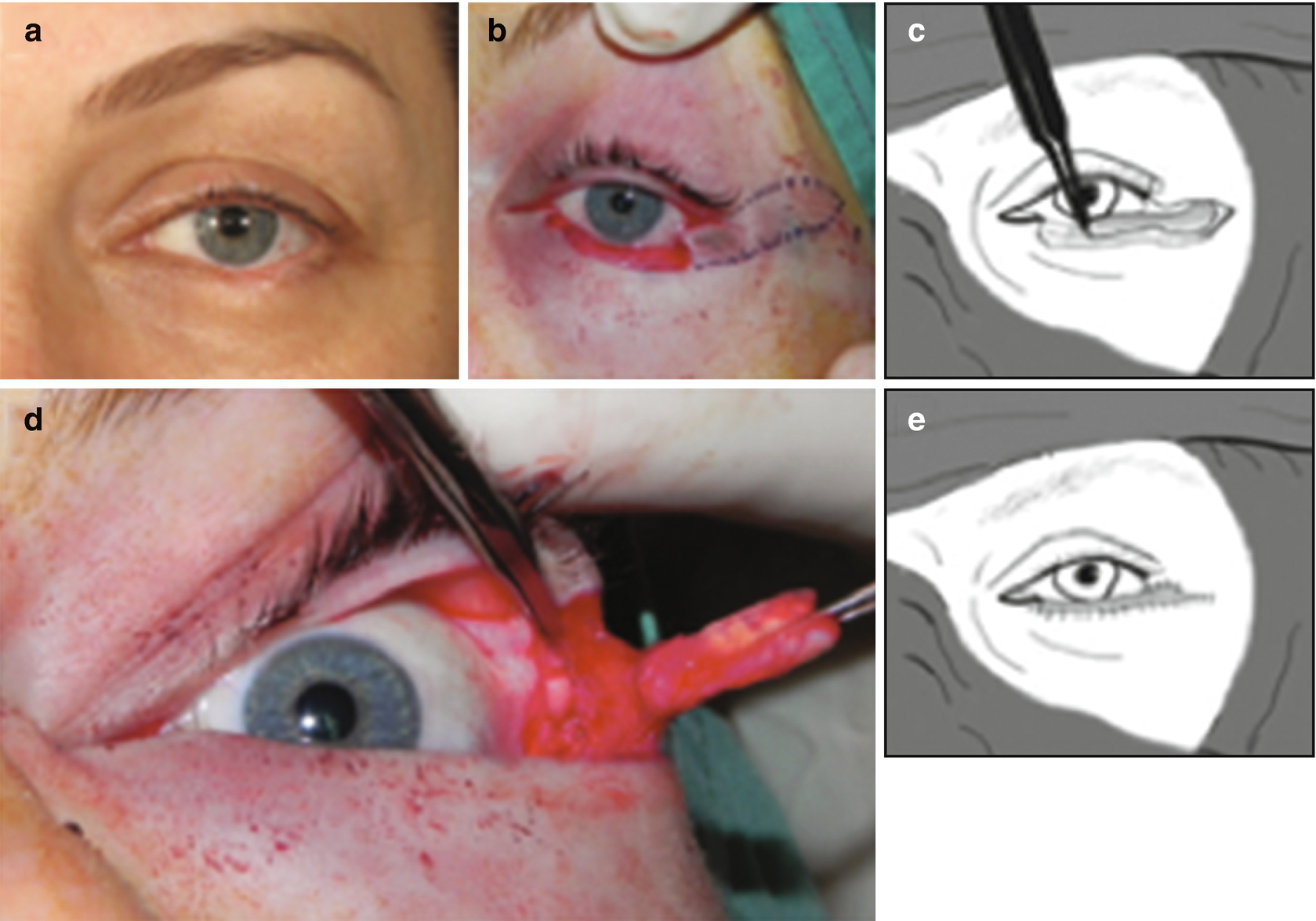
Principles And Techniques Of Eyelid Reconstruction Springerlink

A Cat S Eye With A Conjunctival Pedicle Flap 10 Days After Surgery This Was A Patient Of Dr Cullen S Eyes After Surgery Cats

Observations In Ophthalmology Corneal Opacities In Dogs Cats

Lasik Know The Rewards And The Risks

Corneal Ulcers On Your Dogs Or Cat S Eye
Http Www Eye Vet Com Wp Content Uploads 19 05 Diagnosis And Treatment Of Corneal Ulcerations Pdf

Corneal Ulcers In Animals Wikipedia

Conjunctival Pedicle Grafting Of The Cornea Information

Corneal Endothelial Dystrophy Dog Washington Dc Avo
Corneal Ulcer Treatment Dog Eye Ulcer Treatment In Brisbane

Dry Eye Syndrome Wikipedia
/Prolapsed_gland_of_the_third_eyelid-580285895f9b5805c23baa31.jpeg)
How To Treat Cherry Eye In Dogs

Pin By Cullenwebb Animal Eye Speciali On Eye Diseases Corneal Ulcer Corneal Eyes

Corneal Grafts Davies Veterinary Specialists
Www Agriculturejournals Cz Publicfiles 139 17 Vetmed Pdf
Surgery Of The Eye Veterian Key
Corneal Disease Inherited In Dogs Petmd
Surgery Of The Cornea And Sclera Veterian Key

Superficial Keratectomy And 360º Conjunctival Flap For Bullous Keratopathy In A Dog A Case Report
The Dog House Cornea Repair With Third Eyelid Flap Again
Corneal Lipid
Www Agriculturejournals Cz Publicfiles 139 17 Vetmed Pdf

Conjunctival Pedicle Flap Following Removal Of Corneal Sequestrum From A Cat S Eye Corneal Eyes Cats

Conjunctival Pedicle Flap Used To Prevent This Dog S Eye From Perforating Cullenwebb Corneal Ulcer Dog Eyes Ulcers
03asingapore Dog Eyeball Eye Ulcer Cornea Keratitis Humour Vitreous Condo Internet Advertising Agency Classified Advert For Houses Condos Apartment Advertised By Asiahomes Internet Classified Advertisements Marketing
Why Do Dogs Have A Third Eyelid Healthy Dogs Animal Planet

Deep Corneal Ulceration And Corneal Grafting Procedures

Corneal Epithelial Inclusion Cyst In A Dog

Corneal Grafts Davies Veterinary Specialists

Clinical Case Challenge Blepharospasm Of The Right Eye News Center At Cummings School Of Veterinary Medicine At Tufts University
Surgery Of The Cornea And Sclera Veterian Key
Q Tbn And9gctoywudncljjianeeesklptlrbxqumlkwnxi 4pbj9ur08cdi9h Usqp Cau

Experimental Lamellar Corneal Graft In Dogs Using Preserved Equine Pericardium

Corneal Ulcers In Dogs
Canine Dog Veterinary Surgery Anaesthesiaveterinary Surgery Anaesthesia Singapore Toa Payoh Vets Hamster Medicine Surgery Cases Health Sickness Singapore Singapore Toa Payoh Vets
How To Do Eye Surgery In Deep Corneal Ulcerated Dogs Youtube

South Texas Veterinary Ophthalmology
Q Tbn And9gcsf6urf1qvyyg1bh32hzbldxegtaxugwzffnxqtxcr5dmkxaomr Usqp Cau
Http Centredmv Com Wp Content Uploads 13 11 Fiche Ulcere Corneen Infecte An Pdf

Traumatic Flap Dislocation 10 Years After Lasik Case Report And Literature Review Sciencedirect
Non Healing Superficial Corneal Ulcers In Dogs Mspca Angell

Feline Corneal Sequestrum Mspca Angell

A Modified Technique Of Keratoleptynsis Letter Box For Treatment Of Canine Corneal Edema Associated With Endothelial Dysfunction Giannikaki Veterinary Ophthalmology Wiley Online Library

How To Place A 3rd Eyelid Flap Into A Dog Or Cat Vetgirl Vet Ce Video

Corneal Grafts Davies Veterinary Specialists

Krishikosh Treatment Of Corneal Ulcers With Prp Solid Buffer Combined With Third Eye Lid Flap And Conjunctival Flap In Dogs A Comparative Study

Key Facts Management Of Deep Corneal Ulcers Semantic Scholar
Www Vettimes Co Uk App Uploads Wp Post To Pdf Enhanced Cache 1 Treating Corneal Ulceration In Dogs Part 2 Deep Ulcers Pdf
Corneal Grafts At Animal Eye Care

Conjunctival Pedicle Grafting Of The Cornea Information
Femto Lasik Eye Treatment Lasik Germany

Case Of The Week
Veterinary Conjunctival Flap Graft Animal Eye Consultants
Corneal Ulcer Treatment Dog Eye Ulcer Treatment Seah
Corneal Ulcer Treatment Dog Eye Ulcer Treatment Seah

Cornea Eye Diseases And Disorders Merck Veterinary Manual

Corneal Epithelial Inclusion Cyst In A Dog

Pdf Superficial Keratectomy And 360 Conjunctival Flap For Bullous Keratopathy In A Dog A Case Report

Canine Pedicle Conjunctival Graft Youtube
Dog Cat Ringworm Treatment Thornleigh Veterinary Hospital

Deep Stromal Corneal Ulcers Descemetocele And Iris Prolapse In Animals Emergency Medicine And Critical Care Merck Veterinary Manual

Corneal Endothelial Dystrophy Dog Washington Dc Avo
Uc Davis Ophthalmologists Save Dog S Eyes Following Accident School Of Veterinary Medicine

Conjunctival Flap Cover Surgery 10 Year Review Yao Annals Of Eye Science

Gundersen Flap In Severe Corneal Edema Youtube
Www Vettimes Co Uk App Uploads Wp Post To Pdf Enhanced Cache 1 Treating Corneal Ulceration In Dogs Part 2 Deep Ulcers Pdf

Use Of Porcine Small Intestinal Submucosa For Corneal Reconstruction In Dogs And Cats 106 Cases Goulle 12 Journal Of Small Animal Practice Wiley Online Library
Www Vettimes Co Uk App Uploads Wp Post To Pdf Enhanced Cache 1 Treating Corneal Ulceration In Dogs Part 2 Deep Ulcers Pdf

Corneal Grafts Davies Veterinary Specialists

I Think My Dog S Scratched His Eye What Should I Do Goddard Veterinary Group
1

Healing Of A Corneal Ulcer On A 7 Year Old Mixed Breed Dog The Dog Is Download Scientific Diagram
Www Tieraugen Com Wp Content Uploads 17 09 Kp Pdf
Diagram Of Transpalpebral Third Eyelid Flap Note How The Suture Is Download Scientific Diagram

Veterinary And Travel Stories 2908 Intern Enucleation
Corneal Grafts At Animal Eye Care
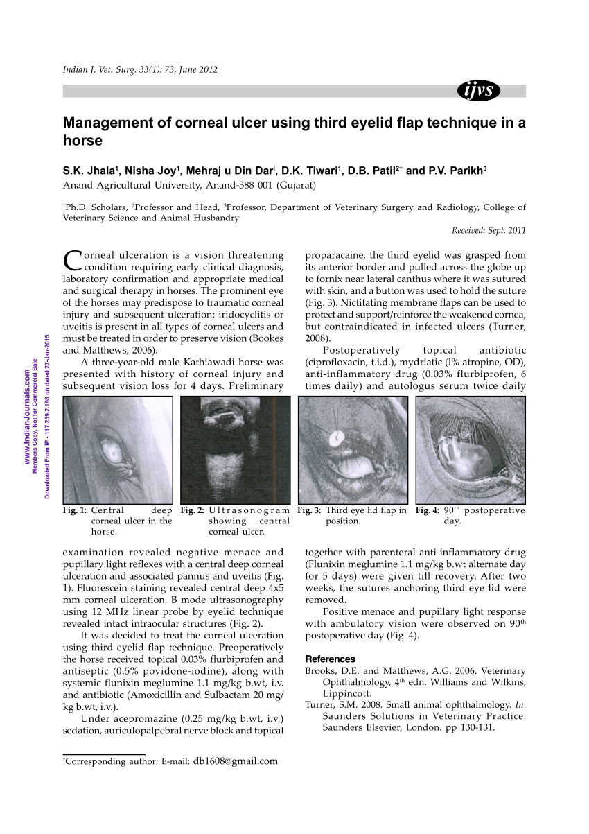
Pdf Management Of Corneal Ulcer Using Third Eyelid Flap Technique In A Horse

My Dog Had A Third Eye Lid Flap Today Around 3 00pm I Picked Her Up St 5 00 She Has The Cone Collar That Is Really


