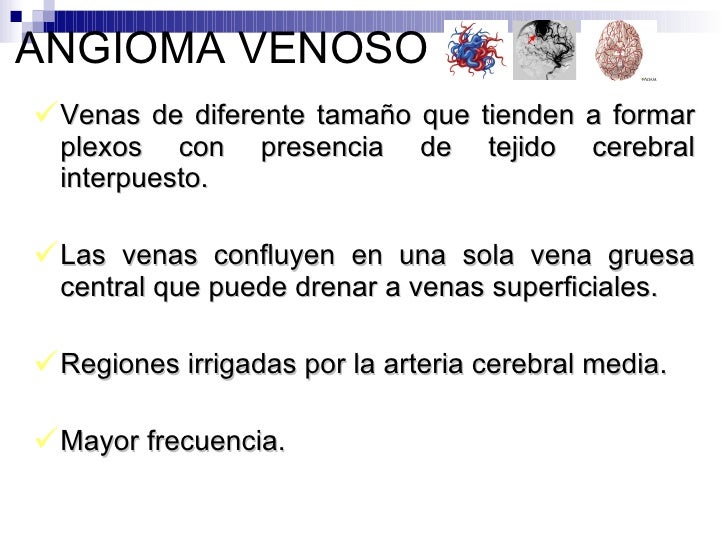
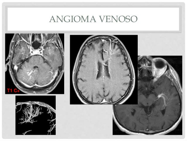
Angioma Venoso Cerebral
Pdfs Semanticscholar Org E284 3bb50bce33bd7cbed2d1ad9b060e173 Pdf

Cavernoma De Cerebelo Unidad De Neurocirugia Rgs

Telangiectasia Capilar Tgc Del Tronco Encefalico Hallazgos En Resonancia Magnetica 1 5 Y 3 Tesla Revista Sintesis
Bjorl Brazilian Journal Of Otorhinolaryngology

Anomalia Venosa Del Desarrollo Intracraneal Asintomatica Sciencedirect

Angioma Venoso Dr Rafael Oliveira
Los angiomas cavernosos comienzan tratándose con corticoides, para reducir la hinchazón Esto pasa, por ejemplo, cuando el angioma tapa la visión u ocluye las vías respiratorias.

Angioma venoso cerebral. La anomalía del desarrollo venoso mientras que el cavernoma permanece sin realce hormonales, causas genéticas, siembra a lo largo de un trade biopsia y las anomalías del desarrollo venoso7 La asociación del cavernoma con las anomalías del desarrollo venoso hay que buscarla, pues su frecuencia puede estar. “Tenía una bomba de relojería en la cabeza” La gráfica descripción que hace Yolanda Lorente (49 años) de su angioma cerebral es una perfecta descripción, nada exagerada, de lo que ha. El angioma senil también se denomina "en guinda", es de color rojo brillante y suele presentarse en el tronco más que en otra partes del cuerpo Es frecuente en personas mayores de 45 años 2.
MR angiography (MRA) was also performed with 3D and 2D timeofflight technique;. Venous angioma of the brain is a dangerous disease, delayed treatment which can lead to unpleasant consequences It is a dilated blood vessels that are intertwined with each other and formed a knot Over time, the blood vessels are «tied» more and more, and this puts pressure on the brain In that case, when the tumour bursts, occurs bleeding. Angiomas are any benign tumors of vascular origin, regardless of which system they represent the circulatory or lymphatic Neoplasms can be located in the superficial layer of cutaneous or mucous membranes, in muscle tissue, in cavities and tissues of internal organs, in the brain.
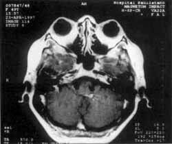
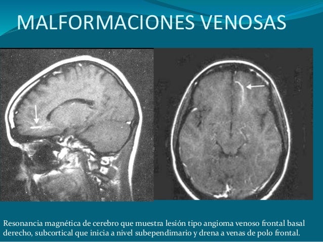
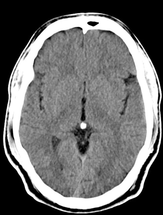
Faktisk er det uden data om cerebral anatomi indsamlet af denne enhed umuligt at detektere anomali, så den kliniske evaluering alene er utilstrækkelig til dens diagnose Men konventionel computertomografi producerer ikke altid de billeder, der er nødvendige for at detektere abnormiteter relateret til venøs angioma, hvorfor det ofte er. Clipping is a handy way to collect important slides you want to go back to later Now customize the name of a clipboard to store your clips. RM cerebral con cortes coronal y axial T1 Gadolinio que muestra (flechas) un angioma venoso, con venas medulares dilatadas en ubicación periventricular frontal derecha, que drenan a una vena.
Únete a Facebook o inicia sesión Correo electrónico o teléfono Contraseña. 5 patients underwent conventional angiography Contrastenhanced MRI demonstrated all the lesions, showing the peripheral. Un angioma cerebral (también denominado hemangioma cerebral) es un tipo de malformación vascular caracterizada por agrupaciones de capilares dilatados Un angioma es una malformación o neoplasia de las células de los vasos sanguíneos que puede aparecer en forma de nódulo o de caverna formada por endotelio que contienen sangre.
If a patient suffers bleeding in the brain and develops a clot related to a venous angioma, it is likely that the patient has a cavernous angioma adjacent to it, notes Neurosurgical Consultants PA When a surgeon finds a cavernous angioma, the surgeon usually treats it by removing the clot and the cavernous angioma, but not disturbing the. A venous angioma is a small abnormal tangle of veins that can occur in the brain Although not technically normal, some people consider a venous angioma, or venous malformation, a normal variant because it occurs fairly frequently (probably in at least a few percent of all people) and because it is rarely associated with any symptoms. Como consecuencia, el angioma venoso se consideraría una causa de la lesión cerebral Otra complicación relacionada con los angiomas venosos es una trombosis de la vena de drenaje Esto puede.
Cerebral Venous Thrombosis Barbara Simons, Geert Lycklama a Nijeholt and Robin Smithuis Radiology department of the Medical Centre Haaglanden in the Hague and the Rijnland hospital in Leiderdorp, the Netherlands Publicationdate Cerebral venous thrombosis is an important cause of stroke especially in children and young adults. Venous angioma of the cerebellum If a brain region in the cerebellum region or an angioma appears in its tissues, several other disturbances appear in the consistency and normal functioning of the organism Venous angioma of the cerebellum provokes such pathological disorders Failure in the consistency of the functioning of skeletal muscles. The angioma vena, Secara teknikal dikenali sebagai anomali perkembangan vena, ia adalah satu set malformasi vaskularIa dianggap sebagai perubahan perkembangan yang dicirikan oleh berterusan pada masa dewasa Keadaan ini biasanya disebabkan oleh perubahan dalam saliran vena semasa peringkat embrio dan menonjol sebagai patologi asimtomatik dengan kursus yang tidak baik.
The authors report 6 cases of cerebral venous angioma and compare the angiographic findings, clinical symptoms, electroencephalographic foci, and histological features to those in 26 previously reported cases They conclude that the socalled venous angioma has little clinical significance, that neither arteries nor capillaries are involved in this type of vascular malformation, and that the small, dilated veins which develop secondarily are due to an abnormality in venous development during. 5 patients underwent conventional angiography Contrastenhanced MRI demonstrated all the lesions, showing the peripheral. O angioma cerebral venoso é uma patologia muito séria que requer diagnóstico precoce e, o mais breve possível Depois de ignorar o problema pode levar a consequências irreparáveis Tratamento medicinal.
Venous angioma of the right hemisphere If we are talking about the hemisphere a layer of gray matter with a thickness of 1345 mm, located on the periphery of the cerebral hemispheres, then the venous angioma of the right hemisphere is fraught with the appearance of such negative symptoms The patient loses the ability to flow smoothly. The patient in this case is a 32yearold man who presented with a clinical history of headache followed by a worsening of his neurological status Neuroimaging studies demonstrated a brain infarct in the posterior fossa, which was related to thrombosis of the draining vein of a cerebral venous angioma. Cerebral venous system can be divided into a superficial and a deep system The superficial system comprises of sagittal sinuses and cortical veins and these drain superficial surfaces of both cerebral hemispheres The deep system comprises of lateral sinus, straight sinus and sigmoid sinus along w.
Introduction Venous angiomas (VA) are benign entities;. The angioma vena, Secara teknikal dikenali sebagai anomali perkembangan vena, ia adalah satu set malformasi vaskularIa dianggap sebagai perubahan perkembangan yang dicirikan oleh berterusan pada masa dewasa Keadaan ini biasanya disebabkan oleh perubahan dalam saliran vena semasa peringkat embrio dan menonjol sebagai patologi asimtomatik dengan kursus yang tidak baik. Venoso O hemangioma venoso é freqüentemente chamado de tumor labial senil ou lago venoso, pois esta formação geralmente ocorre na face dos idosos Especialistas sugerem que o desenvolvimento desses tumores tem um efeito importante na radiação UV.
Angiomas are benign tumors derived from cells of the vascular or lymphatic vessel walls (endothelium) or derived from cells of the tissues surrounding these vessels Angiomas are a frequent occurrence as patients age, but they might be an indicator of systemic problems such as liver disease They are not commonly associated with malignancy. Pedúnculo cerebral, cisterna ambiens y cisterna cuadrigeminal Polígono fetal bilateral con hipoplasia de los segmentos P1 (variante anatómica) Impresión Diagnostica Malformación Vascular del tipo Angioma Venoso (Figuras 1 y 2) Imágenes por Resonancia Magnética Se realizan cortes axiales T1, FLAIR, sagitales T2 apreciándose a nivel de. Clinical Relevance – Cerebral Venous Sinus Thrombosis Cerebral venous sinus thrombosis (CVST) describes the presence of a thrombus within one of the dural venous sinuses The thrombus occludes venous return through the sinuses, and causes an accumulation of deoxygenated blood within the brain parenchyma This in turn can lead to venous infarction The situation is complicated by an accumulation of cerebrospinal fluid, which can no longer drain through the thrombosed venous sinus.
Los angiomas cavernosos comienzan tratándose con corticoides, para reducir la hinchazón Esto pasa, por ejemplo, cuando el angioma tapa la visión u ocluye las vías respiratorias. Angiomas involving cranial nerves are considered rare lesions, radiological characteristics suggest such lesions, although do not define them Definitive diagnosis requires surgical procedure, complete resection in symptomatic patients consist in the best option to avoid worsen of the symptoms and recidivated lesion, knowing that the same. The authors report 6 cases of cerebral venous angioma and compare the angiographic findings, clinical symptoms, electroencephalographic foci, and histological features to those in 26 previously reported cases They conclude that the socalled venous angioma has little clinical significance, that neither arteries nor capillaries are involved in this type of vascular malformation, and that the small, dilated veins which develop secondarily are due to an abnormality in venous development during.
Angioma venoso del cerebro La naturaleza venosa de la enfermedad es una patología menos agresiva, pero dada la localización de la neoplasia, el angioma venoso del cerebro es una enfermedad que no debe ignorarse Debe ser tratado temprano en el desarrollo. If a patient suffers bleeding in the brain and develops a clot related to a venous angioma, it is likely that the patient has a cavernous angioma adjacent to it, notes Neurosurgical Consultants PA When a surgeon finds a cavernous angioma, the surgeon usually treats it by removing the clot and the cavernous angioma, but not disturbing the. Un angioma es una lesión superficial de color rojizo que también se conoce como hemangioma El angioma está formado por pequeños vasos sanguíneos agrupados en una especie de corporación Pueden aparecer en cualquier zona de la superficie de la piel, (en la boca, nariz, vagina o ano) y más raramente pueden afectar a otras partes del cuerpo.
Angioma venoso asociado Finalmente, los angiomas pueden sangrar a través de un punto débil en la pared del angioma en el tejido cerebral circundante Cuanto mayor es el número de angiomas, mayor es la posibilidad de que ocurran una o más hemorragias en algún momento de la vida. However infrequent symptomatic cases may occur Objective Case report and literature review Case report A 6 year old girl was referred with a history of bifrontal, nonpulsatile, headache with no nausea or vomiting Headache intensity was 46/10 The episodes were frequent, 34 times per week. Fig 8 A la izquierda, imagen de un angioma cavernoso a nivel rolándico A la derecha, en otra secuuencia de RM se ve negro el tejido cerebral circundante, por estar embebido de hemosiderina, a consecuencia de microhemorragias previas Fig 9 A la izquierda, RM con angioma cavernoso izquierdo profundo, rodeado de un halo negro de hemosiderina.
Venous angiomas, also called venous malformations, are the brain vascular abnormalities that occur most often Up to 3 percent of the general population may have venous angiomas, notes Riverside Health System Bleeding rarely occurs in venous angiomas, unlike other vascular abnormalities Venous angiomas have enlarged but structurally normal veins, according to Neurosurgical Consultants, PA Surgically removing venous malformations can cause death, because they are typically part of a. Cerebral vascular malformations of smaller and unspecified size have also been described in patients with HHT 510 In its relation to HHT, (caratteristica malattia di RenduOsler), l'altro con emorragie pleuriche e viscerali e angioma venoso cerebrale Pathologica , 1971 Download PDF. A pesar de que es considerada como una malformación venosa del desarrollo, el angioma venoso no resulta exactamente una alteración en el desarrollo cerebral De hecho, esta afección constituye la persistencia en edad adulta de un sistema venoso embrionario, por lo que más que una malformación debería ser considerada como una variante de la normalidad.
Cerebral angiomas are vascular abnormalities comprised of clusters of abnormally dilated blood vessels They can be singular or multiple, and are found in the brain, spinal cord, and rarely, in other areas of the body including the skin and retina They are also known as cavernous angioma cavernous hemangioma cavernous venous malformation. Un angioma es una lesión superficial de color rojizo que también se conoce como hemangioma El angioma está formado por pequeños vasos sanguíneos agrupados en una especie de corporación Pueden aparecer en cualquier zona de la superficie de la piel, (en la boca, nariz, vagina o ano) y más raramente pueden afectar a otras partes del cuerpo. II Departamentos de Lesado Cerebral – Rede Sarah de Hospitais de Reabilitação, Fortaleza CE, Brazil Developmental venous anomaly (DVA), also known as venous angioma, is becoming the most commonly encountered intracranial vascular malformation in central nervous system (CNS) imaging 1,2.
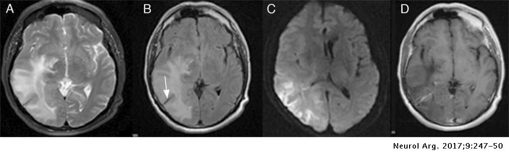
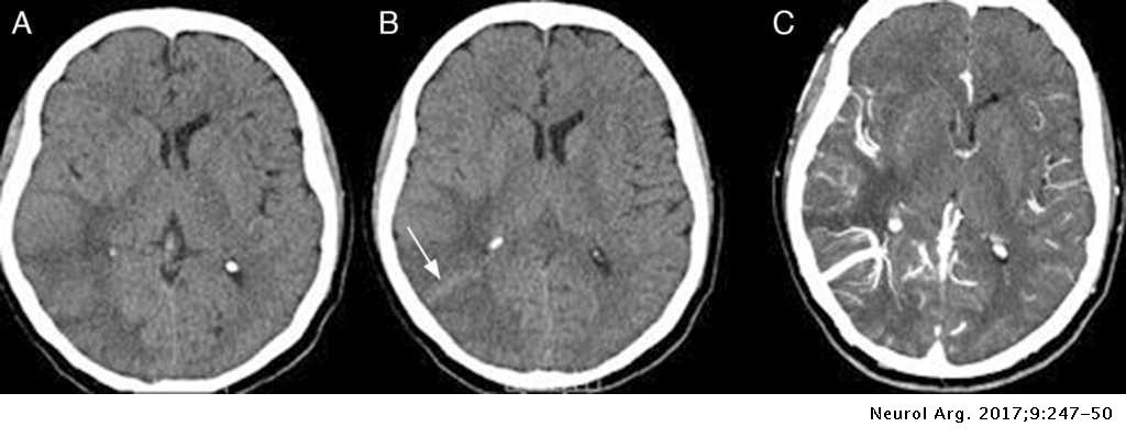
Phenomenon in the pathophysiology of cerebral venous thrombosis (CVT), caused by congestion of vessels that leads to decreased cerebral blood flow7 Thus, decreased ADC in abnormal brain areas associated with CVT may not have the same prognostic value as those associated with pure arterial stroke Clinical improvement in our patient. You just clipped your first slide!. Abstract Several unusual cases of cerebral venous angiomas as well as some characteristic cases are reported The characteristic angiographic feature is that of a collection of dilated medullary veins draining into a single large draining vein, which appears first in the early venous phase and persists into the late venous phase of the arteriogram.
Cerebral venous angioma (developmental venous anomaly) is the most common cerebral vascular malformation It consists of an intraparenchymal tangle/cluster of dilated medullary veins converging on a single enlarged draining vein It rarely bleeds Differential diagnosis vascular malformation , venous varix. We studied 17 patients with venous angiomas, 4 of whom had associated cavernous angiomas All underwent MRI with spinecho T1 and T2weighted images and T1weighted images after GdDTPA;. Developmental venous anomaly (DVA), also known as cerebral venous angioma, is a congenital malformation of veins which drain normal brain They were thought to be rare before crosssectional imaging but are now recognized as being the most common cerebral vascular malformation, accounting for ~55% of all such lesions.
Cavernous angiomas of theer m er m h h cranial nerves surgical management,l ca a er g results, and complicationsl s com i sl s com i s ABSTRACTR T Introduction o u i Cavernous brain malformations (cavernous angiomas), cavernomas, as well as occulted angiographic vascular malformations presents 03 – 05%. Un hemangioma se puede presentar con otras afecciones poco frecuentes Se pueden realizar otros exámenes para buscar problemas relacionados Tratamiento La mayoría de los hemangiomas pequeños o sin complicaciones puede no necesitar tratamiento Con frecuencia desaparecen por sí mismos y la apariencia de la piel regresa a la normalidad. The patient in this case is a 32yearold man who presented with a clinical history of headache followed by a worsening of his neurological status Neuroimaging studies demonstrated a brain infarct in the posterior fossa, which was related to thrombosis of the draining vein of a cerebral venous angioma.
Venous angioma of the brain is a dangerous disease, delayed treatment which can lead to unpleasant consequences It is a dilated blood vessels that are intertwined with each other and formed a knot Over time, the blood vessels are «tied» more and more, and this puts pressure on the brain In that case, when the tumour bursts, occurs bleeding. MR angiography (MRA) was also performed with 3D and 2D timeofflight technique;. Gli autori descrivono, otto casi di angioma venoso (AV) esaminati con RM ed angiografia a risonanza magnetica con tecnica 2D TOF, sequenze FISPLa buona qualità delle immagini ottenute, ha permesso.
El angioma senil también se denomina "en guinda", es de color rojo brillante y suele presentarse en el tronco más que en otra partes del cuerpo Es frecuente en personas mayores de 45 años 2. Angiomas venosos estão frequentemente associados a angiomas cavernosos Não há indicação cirúrgica de angioma venoso, pois sua remoção pode resultar em infarto venoso grave12 Nessa série de 9 pacientes, a ressecção do angioma cavernoso foi total em todos os casos. Venous angiomas are vascular malformations diagnosed with increasing frequency in the last few years, due to the availability of new neuroimaging techniqueso In this paper we describe a case of venous angioma studied with cerebral angiography, CT and MRI and the literature in relation with this specific vascular lesiono.
Hemangioma na retina Conforme o angioma cresce, as paredes dos vasos sanguíneos podem debilitarse até ao ponto de se romperem, causando danos nos tecidos próximos Os derrames dos angiomas da retina podem interferir com a visão Para manter a visão saudável é muito importante detectar o angioma o mais cedo possível e acompanhálo de. Un hemangioma se puede presentar con otras afecciones poco frecuentes Se pueden realizar otros exámenes para buscar problemas relacionados Tratamiento La mayoría de los hemangiomas pequeños o sin complicaciones puede no necesitar tratamiento Con frecuencia desaparecen por sí mismos y la apariencia de la piel regresa a la normalidad. Detta faktum gör venules hänvisar till venous angioma förvärva ett maneter utseende och få namnet Caput medusae Venös anomali kan hittas i någon region i hjärnan, men ligger oftast i frontalloberna i hjärnbarken och den bakre fossa På samma sätt är två tredjedelar av de totala venösa angioma som hittills hittats i cerebellum.
Embora seja considerado uma malformação venosa do desenvolvimento, o angioma venoso não é exatamente uma alteração no desenvolvimento cerebral De fato, essa condição constitui a persistência na vida adulta de um sistema venoso embrionário, e é por isso que mais do que uma malformação deve ser considerada uma variante da normalidade.

Angioma Venoso Asociado A Hemorragia Cerebelosa Presentacion De Un Caso Sciencedirect

Questions Answered Angioma Alliance

Cerebral Venous Angioma Radiology Case Radiopaedia Org

Malformaciones Vasculares Cerebrales
Http Pdf Posterng Netkey At Download Index Php Module Get Pdf By Id Poster Id 1112

Radio Medic Angioma Venoso Frontal Derecho Con La Facebook
Www Neurocirugiachile Org Pdfrevista V45 N3 19 Chacon P250 V45n3 19 Pdf

Cerebral Venous Angioma Of The Pons Complicated By Nonhemorrhagic Infarction In Journal Of Neurosurgery Volume 101 Issue 4 04
Www Elsevier Es Index Php P Revista Prevista Pdf Simple Pii S R 8

Eduimagen Eduimagen Agrego 4 Fotos Nuevas Al Album Facebook

Signo De La Cabeza De Medusa

Blog Helpsaude Malformacoes Vasculares Do Cerebro
Anomalias Venosas Del Desarrollo Angiomas Venosos Neurowikia
Infarto Cerebral Secundario A Anomalia Del Desarrollo Venoso En Un Paciente Oncologico Neurologia Argentina
Angioma Venoso Frontal Que Es Un Angioma Se Puede Eliminar Vena Colapsada En Los Sintomas Del Brazo
Malformaciones Vasculares

Investigation Of Thunderclap Headache In Cavernous Angioma When Magnetic Resonance Makes The Difference Case Report
Radiologia Brasileira Sinais Em Neurorradiologia Parte 1

Malformaciones Vasculares Cerebrales Tema 9 Parte Ii Unidad De Neurocirugia Rgs
Malformaciones Vasculares Cerebrales
Bjorl Brazilian Journal Of Otorhinolaryngology

Cavernous Hemangioma Wikipedia

Hemorragia Intracerebral Secundaria A Trombosis Espontanea De Una Anomalia Venosa Del Desarrollo 2 Casos Y Revision De La Literatura

Telangiectasia Capilar Tgc Del Tronco Encefalico Hallazgos En Resonancia Magnetica 1 5 Y 3 Tesla Revista Sintesis
Malformaciones Vasculares Cerebrales

Epos S 0919

Malformaciones Vasculares Cerebrales Tema 9 Parte Ii Unidad De Neurocirugia Rgs
Infarto Cerebral Secundario A Anomalia Del Desarrollo Venoso En Un Paciente Oncologico Neurologia Argentina
Developmental Venous Anomaly Radiology Case Radiopaedia Org

Unp1pa1efy Hm

Cerebral Venous Angioma Of The Pons Complicated By Nonhemorrhagic Infarction In Journal Of Neurosurgery Volume 101 Issue 4 04

Developmental Venous Anomaly Radiology Reference Article Radiopaedia Org

Que Es Un Cavernoma Parte 1 Concepto Unidad De Neurocirugia Rgs
Http Www Webcir Org Revistavirtual Articulos 15 Marzo Espana Cavernomas Ing Pdf

Thrombosed Developmental Venous Anomaly Associated With Cerebral Venous Infarct

Cavernoma Es Necesario Operar Neurovist El Blog

Angioma Venoso Asociado A Hemorragia Cerebelosa Presentacion De Un Caso Sciencedirect

Malformacoes Vasculares Tipos E Diagnostico
Angiomas Arteriovenosos Do Cerebro

Hemorragia Intracerebral Secundaria A Trombosis Espontanea De Una Anomalia Venosa Del Desarrollo 2 Casos Y Revision De La Literatura

Infarto Cerebral Secundario A Anomalia Del Desarrollo Venoso En Un Paciente Oncologico Neurologia Argentina
Infarto Cerebral Secundario A Anomalia Del Desarrollo Venoso En Un Paciente Oncologico Neurologia Argentina
Q Tbn And9gcqesf3d6zgi8aoglhx5xcfx Wdeba5 Yzk4fa Pppc Usqp Cau
Http Pdf Posterng Netkey At Download Index Php Module Get Pdf By Id Poster Id 1112

Epos Trade

Hemorragia Subaracnoidea No Aneurismatica
1

Cerebral Venous Angioma Of The Pons Complicated By Nonhemorrhagic Infarction In Journal Of Neurosurgery Volume 101 Issue 4 04

Cavernomas O Angiomas Cavernosos Neurowikia
Asociacion Entre Angioma Cavernoso Y Glioma Cerebral Reporte De Dos Casos Y Revision De La Literatura Acerca De Los Llamados Angiogliomas

Que Es Un Cavernoma Parte 1 Concepto Unidad De Neurocirugia Rgs

Hemorragia Intracerebral Secundaria A Trombosis Espontanea De Una Anomalia Venosa Del Desarrollo 2 Casos Y Revision De La Literatura

Angioma Venoso Asociado A Hemorragia Cerebelosa Presentacion De Un Caso Sciencedirect
Q Tbn And9gcsqnywaea95v6tnquc0rpich1gwkv21a26yqaspkybw25tehb N Usqp Cau

Cirurgia Estereotaxica Guiada Para Angiomas Cavernosos
Www Angioma Org Wp Content Uploads 06 Patientbrochure18spanish Pdf

Investigation Of Thunderclap Headache In Cavernous Angioma When Magnetic Resonance Makes The Difference Case Report

Signo De La Cabeza De Medusa
Www Neurocirugiachile Org Pdfrevista V45 N3 19 Chacon P250 V45n3 19 Pdf

Anomalia De Desenvolvimento Venoso E Meningioma Youtube
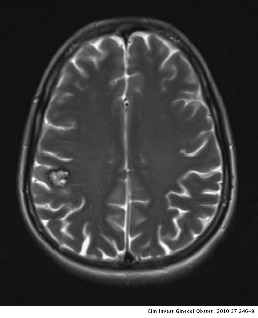
Diagnostico De Cavernoma Cerebral Durante El Embarazo Clinica E Investigacion En Ginecologia Y Obstetricia

Nuestra Experiencia En El Manejo De Los Cavernomas De Tronco De Talamo Y De Ganglios Basales Revista Argentina De Neurocirugia

Rm Do Cranio Anomalia Do Desenvolvimento Venoso Youtube

Que Es Un Cavernoma Parte 1 Concepto Unidad De Neurocirugia Rgs

Cerebral Venous Angioma Of The Pons Complicated By Nonhemorrhagic Infarction In Journal Of Neurosurgery Volume 101 Issue 4 04

Que Es Un Cavernoma Parte 1 Concepto Unidad De Neurocirugia Rgs
O Que E Angioma Venoso Sintomas E Tratamento Tua Saude

Angioma Venoso Causas Sintomas Diagnostico Tratamento Competentemente Sobre Saude No Ilive
Www Neurocirugiachile Org Wp Content Uploads 18 11 Elizondo P171 V44n2 18 Pdf

O Que E Angioma Venoso Sintomas E Tratamento Tua Saude

Malformaciones Vasculares Cerebrales

Sindrome De Sturge Weber Variabilidad Clinica Y De Neuroimagen Anales De Pediatria

Developmental Venous Anomaly Pons Radiology Case Radiopaedia Org
Cerebral Venous Angioma Radiology Case Radiopaedia Org
Angioma Cavernoso Cerebral Gigante En Paciente Menor De Un Ano Descripcion De Un Caso
Http Www Webcir Org Revistavirtual Articulos 15 Marzo Espana Cavernomas Ing Pdf

Malformaciones Vasculares Cerebrales
Blog Helpsaude Malformacoes Vasculares Do Cerebro

Signo De La Cabeza De Medusa

Resonancia Magnetica Craneal En Secuencia T2 Donde Se Muestra Imagen Download Scientific Diagram

Pdf Ct Of Venous Angiomas Of The Brain Semantic Scholar

O Que E Angioma Venoso Sintomas E Tratamento Tua Saude
Www Angioma Org Wp Content Uploads 06 Patientbrochure18spanish Pdf

Angioma Cavernoso

Malformaciones Vasculares Cerebrales Tema 9 Parte Ii Unidad De Neurocirugia Rgs

Figure 2 From Treatment Differences In Cases With Venous Angioma Semantic Scholar

Angioma Cerebral Como Nos Afecta Mejor Con Salud

Angiomas Arteriovenosos Do Cerebro
Http Www Webcir Org Revistavirtual Articulos 15 Marzo Espana Cavernomas Ing Pdf
Http Www Webcir Org Revistavirtual Articulos 15 Marzo Espana Cavernomas Ing Pdf
1
Www Redalyc Org Pdf 35 Pdf

Malformaciones Vasculares Cerebrales
Www Medigraphic Com Pdfs Revcubmed Cm 19 Cm192h Pdf

Angioma Venoso Caracteristicas Sintomas Y Tratamientos

Que Es Un Angioma Venoso Mejor Con Salud

Angiomas Venosos Y Cefalea En Pediatria Un Caso Clinico

Flickriver La Neuroimagen S Most Interesting Photos


